WB result of CD96 Recombinant Rabbit mAb
Primary antibody: CD96 Recombinant Rabbit mAb at 1/1000 dilution
Lane 1: Daudi whole cell lysate 20 µg
Lane 2: MCF7 whole cell lysate 20 µg
Lane 3: A375 whole cell lysate 20 µg
Lane 4: Molt-4 whole cell lysate 20 µg
Lane 5: HuT-78 whole cell lysate 20 µg
Negative control: Daudi whole cell lysate; MCF7 whole cell lysate
Secondary antibody: Goat Anti- rabbit IgG, (H+L), HRP conjugated at 1/10000 dilution
Predicted MW: 66 kDa
Observed MW: 100, 160 kDa
This blot was developed with high sensitivity substrate
CD96 Recombinant Rabbit mAb (S-5322)
CD96 Recombinant Rabbit mAb (S-5322)
Price:
Regular price
$100 USD
Regular price
Sale price
$100 USD
Unit price
per
For shipping services or bulk orders, you may request a quotation.
Secure checkout with

View full details
Product Details
Product Details
Product Specification
| Host | Rabbit |
| Antigen | CD96 |
| Synonyms | T-cell surface protein tactile; Cell surface antigen CD96; T cell-activated increased late expression protein |
| Location | Membrane |
| Accession | P40200 |
| Clone Number | S-5322 |
| Antibody Type | Recombinant mAb |
| Isotype | IgG |
| Application | WB, IHC-P |
| Reactivity | Hu |
| Positive Sample | A375, Molt-4, HuT-78 |
| Purification | Protein A |
| Concentration | 0.5 mg/ml |
| Conjugation | Unconjugated |
| Physical Appearance | Liquid |
| Storage Buffer | PBS, 40% Glycerol, 0.05% BSA, 0.02% sodium azide |
| Stability & Storage | 12 months from date of receipt / reconstitution, -20 °C as supplied |
Dilution
| application | dilution | species |
| WB | 1:1000 | Hu |
| IHC-P | 1:100 | Hu |
Background
CD96, also known as TACTILE (T cell activation, late expression protein), is a type I transmembrane glycoprotein belonging to the immunoglobulin superfamily, primarily expressed on the surface of activated T cells and natural killer (NK) cells. The extracellular region of this protein contains immunoglobulin-like domains, and through binding to PVR (poliovirus receptor, CD155) on the surface of target cells, it mediates the adhesion between NK cells and target cells, playing an important role in cell-cell adhesion and signal regulation during the late phase of immune responses. Functionally, CD96 is now widely recognized as a novel inhibitory immune checkpoint. It negatively regulates the activation and cytotoxic functions of NK cells and CD8+ T cells by competing with the costimulatory receptor CD226 for binding to their shared ligand CD155, with higher affinity than CD226. Studies have shown that in various malignancies such as acute myeloid leukemia and hepatocellular carcinoma, CD96 expression on NK cells is significantly upregulated and is closely associated with dysfunction induced by immunosuppressive factors such as TGF-β1. NK cells with high CD96 expression exhibit reduced IFN-γ secretion and decreased cytotoxic activity, indicating a poor clinical prognosis. Given its critical role in tumor immune evasion, blocking the CD96-CD155 interaction has emerged as a novel strategy to enhance anti-tumor immune responses. CD96 is actively being investigated as a next-generation immune checkpoint target, offering new directions for improving the efficacy of immunotherapies.
Picture
Picture
Western Blot
Immunohistochemistry
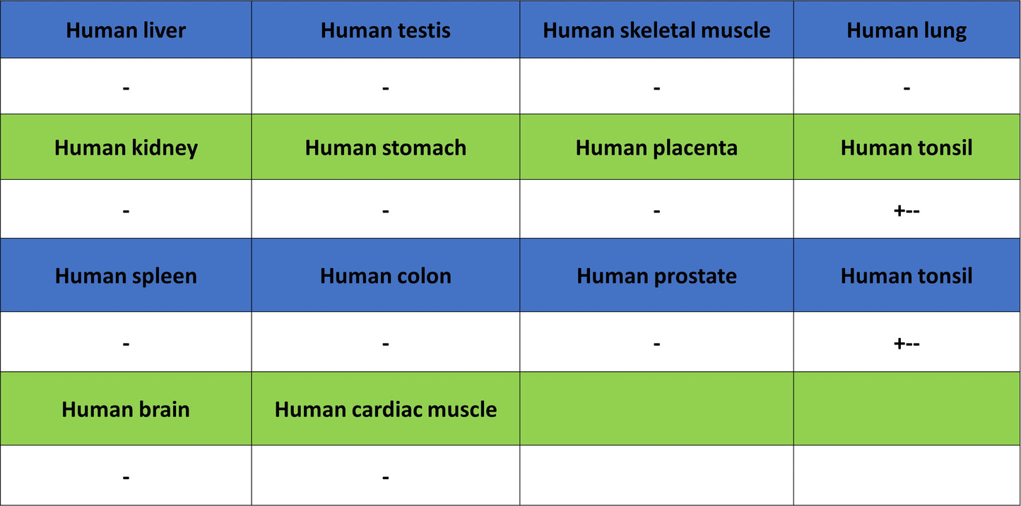
IHC shows positive staining in paraffin-embedded human cervical squamous cell carcinoma. Anti-CD96 antibody was used at 1/100 dilution, followed by a HRP Polymer for Rabbit IgG (ready to use). Counterstained with hematoxylin. Heat mediated antigen retrieval with Tris/EDTA buffer pH9.0 was performed before commencing with IHC staining protocol.
IHC shows positive staining in paraffin-embedded human NK/T-cell lymphoma. Anti-CD96 antibody was used at 1/100 dilution, followed by a HRP Polymer for Rabbit IgG (ready to use). Counterstained with hematoxylin. Heat mediated antigen retrieval with Tris/EDTA buffer pH9.0 was performed before commencing with IHC staining protocol.