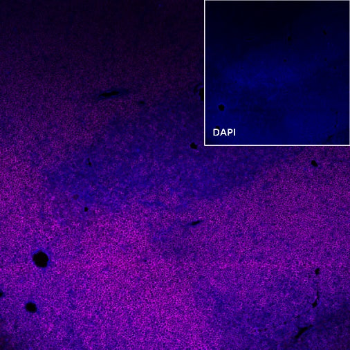
IF shows positive staining in paraffin-embedded mouse thymus. Anti- CD8α (Alexa Fluor® 647 Conjugate) antibody was used at 1/200 dilution (magenta). Counterstained with DAPI (Blue). Heat mediated antigen retrieval with Tris/EDTA buffer pH9.0 was performed before commencing with IF staining protocol.
CD8α Recombinant Rabbit mAb (Alexa Fluor® 647 Conjugate) (SDT-142-52)
CD8α Recombinant Rabbit mAb (Alexa Fluor® 647 Conjugate) (SDT-142-52)
Price:
Regular price
$180 USD
Regular price
Sale price
$180 USD
Unit price
per
For shipping services or bulk orders, you may request a quotation.
Secure checkout with

View full details
Product Details
Product Details
Product Specification
| Host | Rabbit |
| Antigen | CD8α |
| Synonyms | Lyt-2 |
| Immunogen | Synthetic Peptide |
| Location | Cell membrane |
| Accession | P01731 |
| Clone Number | SDT-142-52 |
| Antibody Type | Recombinant mAb |
| Application | IF |
| Reactivity | Ms |
| Purification | Protein A |
| Concentration | 2 mg/ml |
| Conjugation | Alexa Fluor® 647 |
| Physical Appearance | Liquid |
| Storage Buffer | PBS, 1% BSA, 0.3% Proclin 300 |
| Stability & Storage | 12 months from date of receipt / reconstitution, 2 to 8 °C as supplied. |
Dilution
| application | dilution | species |
| IF | 1:200 |
Background
CD8α encodes the CD8 alpha chain of the αβT cells, proposed as a quantifiable indicator for CD8+ CTL recruitment or activity assessments and a robust biomarker for responses to anti-PD-1/PD-L1 therapy. In NK-cells, the presence of CD8A homodimers at the cell surface provides a survival mechanism allowing conjugation and lysis of multiple target cells. CD8A homodimer molecules also promote the survival and differentiation of activated lymphocytes into memory CD8 T-cells.
Picture
Picture
Immunofluorescence