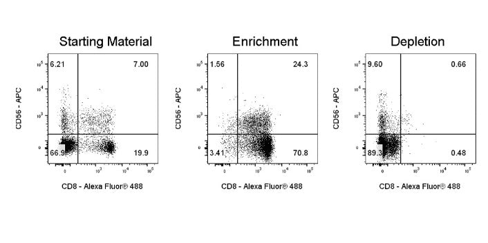
CD8+ T cells are enriched or depleted from human PBMCs using CD8 Nanobeads, S or L Separation Columns, and a magnetic separator. Subsequently, fluorescent staining is performed with anti-CD8 (clone: OKT8) Alexa Fluor® 488, and the live cell population is gated for analysis.
CD8 Nanobeads, human(RUO)
CD8 Nanobeads, human(RUO)
Price:
Regular price
$300 USD
Regular price
Sale price
$300 USD
Unit price
per
For shipping services or bulk orders, you may request a quotation.
Secure checkout with

View full details
Product Details
Product Details
Product Specification
| Format | Dextran-coated iron tetroxide nanomagnetic beads coupled with an anti-human CD8 monoclonal antibody |
| Capacity |
2 mL magnetic beads: suitable for a total cell count of 1×10⁹, and can perform up to 100 separations 1 mL magnetic beads: suitable for 5×10⁸total cells, and can perform up to 50 separations |
| Principle of Separation | CD8⁺ cells are magnetically labeled using CD8 Nanobeads for humans. The cell suspension is then loaded into a separation column placed within the sorter's magnetic field. The magnetically labeled CD8⁺ cells remain retained in the column, while the unlabeled cells elute; this fraction constitutes the negative fraction after removal of CD8⁺ cells. After removing the separation column from the magnetic field, the magnetically captured CD8⁺ cells can be eluted, yielding the positive sorted fraction. If higher purity is required, the positive sorted fraction containing CD8⁺ cells must be further separated using a second column. |
| Reactivity | Human |
| Magnetic Bead Size | 20 nm |
| Separation Method | Column-Based |
| Selection Strategy | Positive Selection |
| Stability & Storage | Store protected from light at 2–8°C; do not freeze. |
Background
CD8 antigen forms a complex with the T-cell receptor and serves as a co-molecule on CD8+ cytotoxic T cells, assisting the TCR heterodimer in recognizing MHC class I–peptide complexes. The CD8 molecule consists of an α/β heterodimer or an α/α homodimer. It is highly expressed on cytotoxic T cells and weakly expressed on some NK cells. CD8 is present on most thymocytes and approximately one-third of peripheral blood T cells. CD8+ cytotoxic T cells play a crucial role in eliminating virus-infected cells and tumor cells.
Protocol
Note: This kit does not include the sorting buffer; it must be provided separately. The corresponding Stater catalog number isS0D3018
Taking S as an exampleSeparation Columnfor instance
Steps |
Operating procedures |
Dosage / Time |
|
Magnetic labeling |
1 |
Count the total number of cells |
— |
2 |
Centrifuge the cell suspension at 400×g for 7 minutes, and completely discard the supernatant |
400×g, 7 min |
|
3 |
Resuspend every 1×10⁷ cells in 80 μL of buffer Note:Adjust the volume of the buffer proportionally according to the sample volume |
Resuspend the cells |
|
4 |
Add 20 μL of CD8 NanoBeads per 1×10⁷ cells Note:When the number of cells increases, increase the volumes of all reagents proportionally |
Add 20 μL per 1×10⁷ cells |
|
5 |
Mix thoroughly and incubate in the dark at 2–8℃ for 15 minutes |
Incubate for 15 minutes |
|
6 |
Wash every 1×10⁷ cells with 1 mL of buffer, centrifuge at 400×g for 7 minutes, and completely discard the supernatant. |
Add 1 mL to 1×10⁷ cells |
|
7 |
Resuspend up to 1×10⁸ cells in 500 μL of buffer8Cells |
Resuspend cells |
|
Magnetic separation |
8 |
Place the S-type separation column into the magnetic field of the corresponding magnetic separator, and rinse the column with 500 μL of buffer. |
Rinse the separation column |
9 |
Load the cell suspension onto the separation column and collect the flow-through (unlabeled cells). |
— |
|
10 |
Wash the column three times with 500 μL of buffer each time, adding the next wash only after the liquid inside the column has completely drained. |
3 × 500 μL of buffer |
|
11 |
Combine all flow-through fractions into the negative fraction; remove the separation column from the magnetic field and place it over the collection tube. |
— |
|
12 |
Add 1 mL of buffer to the column, and quickly and firmly push the accompanying plunger to elute the magnetically labeled positive cells. Note:For higher purity, use a new column and repeat the separation once. |
Collect the target cells. |
With LSeparation Columnas an example
Steps |
Operating method |
Dosage / Time |
|
Magnetic labeling |
1 |
Count the total number of cells |
— |
2 |
Centrifuge the cell suspension at 400×g for 7 minutes, and completely discard the supernatant. |
400×g, 7 min |
|
3 |
Resuspend every 1×10⁷ cells in 80 μL of buffer. Note:Adjust the volume of the buffer proportionally according to the sample volume. |
Resuspend the cells. |
|
4 |
Add 20 μL of CD8 NanoBeads per 1×10⁷ cells. Note:When the cell quantity increases, increase the volume of all reagents proportionally at the same time. |
Add 20 μL per 1×10⁷ cells. |
|
5 |
Mix thoroughly and incubate in the dark at 2–8℃ for 15 minutes. |
Incubate for 15 min. |
|
6 |
Wash every 1×10⁷ cells with 1 mL of buffer, centrifuge at 400×g for 7 minutes, and completely discard the supernatant. |
Add 1 mL to 1×10⁷ cells |
|
7 |
Resuspend up to 1×10 in 500 μL of buffer8Cells |
Resuspend cells |
|
Magnetic separation |
8 |
Place the L-shaped separation column into the magnetic field of the corresponding magnetic separator, and rinse the column with 3 mL of buffer. |
Rinse the separation column |
9 |
Load the cell suspension onto the separation column and collect the effluent (unlabeled cells). |
— |
|
10 |
Wash the column with buffer three times, each time using 3 mL; add the next wash only after the liquid inside the column has completely drained. |
3 × 3 mL of buffer |
|
11 |
Combine all effluents into the negative fraction; remove the separation column from the magnetic field and place it over the collection tube. |
— |
|
12 |
Add 5 mL of buffer to the column, and quickly and firmly push the accompanying plunger to elute the magnetically labeled positive cells. Note:For higher purity, use a new column and repeat the separation once. |
Collect the target cells. |
Picture
Picture
Validation Data