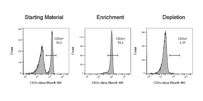
Using the CD3ε Nanobeads Kit, mouse S(D) separation columns, and a sorter, CD3ε-positive cells were isolated from the single-cell suspension. The cells were fluorescently labeled with an anti-CD3ε+ (clone: S-R491) Alexa Fluor® 488 antibody (SDT Cat: S0B5473), and the "live cell+" population was selected.
CD3ε Nanobeads Kit, mouse (RUO)
CD3ε Nanobeads Kit, mouse (RUO)
Price:
Regular price
$335 USD
Regular price
Sale price
$335 USD
Unit price
per
For shipping services or bulk orders, you may request a quotation.
Secure checkout with

View full details
Product Details
Product Details
Product Specification
| Format | Biotinylated anti-mouse CD3ε monoclonal antibody and streptavidin-conjugated nanomagnetic beads, in a PBS buffer system containing BSA and Poloxamer 188. |
| Capacity |
1 mL magnetic beads: Suitable for a total cell count of 5×10⁸, allowing for up to 50 sorting runs. 2 mL magnetic beads: Suitable for a total cell count of 1×10⁹, allowing for up to 100 sorting runs. |
| Principle of Separation | First, CD3ε+ cells are labeled with a CD3ε-biotin–conjugated primary antibody. Subsequently, the cells are magnetically labeled using biotin-conjugated nanomagnetic beads. The cell suspension is then placed in a separation column within a magnetic field. The CD3ε+ cells that have been labeled with the magnetic beads remain retained in the column, while the unlabeled cells flow through; thus, CD3ε+ cells are separated from the other cell populations. |
| Reactivity | Mouse |
| Magnetic Bead Size | 20 nm |
| Separation Method | Column-Based |
| Selection Strategy | Positive Selection |
| Stability & Storage | Store protected from light at 2–8 °C; do not freeze. |
Background
CD3ε is a transmembrane protein with a molecular weight of 20 kDa and serves as a subunit of the T-cell receptor (TCR) complex. Together with the γ and δ subunits of CD3, it plays a crucial role in the assembly and expression of the TCR complex. CD3 is expressed on the surface of thymocytes, mature T lymphocytes, and natural killer T cells (NKT cells). Using the CD3ε nanosphere kit, mouse T lymphocytes and NKT cells can be depleted from various tissues, including the spleen, thymus, and lymph nodes.
Picture
Picture
Validation Data