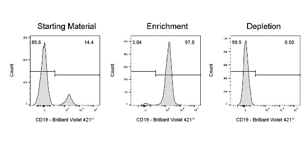
Enrich or deplete CD19+ cells from human PBMCs (human peripheral blood mononuclear cells). Subsequently, perform fluorescent staining with anti-CD19 (clone: HIB19) Brilliant Violet 421™ and gate on the live cell population for analysis.
CD19 Nanobeads, human(RUO)
CD19 Nanobeads, human(RUO)
Price:
Regular price
$285 USD
Regular price
Sale price
$285 USD
Unit price
per
For shipping services or bulk orders, you may request a quotation.
Secure checkout with

View full details
Product Details
Product Details
Product Specification
| Format | Dextran-coated iron tetroxide magnetic nanoparticles conjugated with an anti-human CD19 monoclonal antibody |
| Capacity |
2 mL magnetic beads: suitable for a total cell count of 1×10⁹, and can perform up to 100 separations 1 mL magnetic beads: suitable for 5×10⁸total cells, and can perform up to 50 separations |
| Principle of Separation | CD19⁺ cells are magnetically labeled using CD19 Nanobeads. The cell suspension is then loaded into a separation column placed within the sorter's magnetic field. The magnetically labeled CD19⁺ cells remain retained in the column, while the unlabeled cells elute; this fraction constitutes the negative fraction after depletion of CD19⁺ cells. After removing the separation column from the magnetic field, the magnetically captured CD19⁺ cells can be eluted, yielding the positive sorted fraction. To further enhance purity, the positive fraction containing CD19⁺ cells should be subjected to a second separation column. |
| Reactivity | Human |
| Magnetic Bead Size | 20 nm |
| Separation Method | Column-Based |
| Selection Strategy | Positive Selection |
| Stability & Storage | Store protected from light at 2–8°C; do not freeze. |
Background
CD19 Nanobeads are designed to isolate human B cells based on the expression of the CD19 antigen. CD19 is a 95–120 kDa glycosylated transmembrane protein that plays a crucial role in the signal transduction pathways regulating B lymphocyte development, activation, and differentiation. CD19 is expressed on cells of the B-cell lineage, from the early committed pro-B cell stage through the pre-B cell stage, including most malignant B cells. Its expression is downregulated during differentiation into plasma cells. In addition, CD19 is also expressed on follicular dendritic cells.
Protocol
Note: This kit does not include sorting buffer; please prepare it yourself. The corresponding Stater catalog number isS0D3018
Taking S as an exampleSeparation ColumnFor instance
Steps |
Operating procedures |
Dosage / Time |
|
Magnetic labeling |
1 |
Count the total number of cells |
— |
2 |
Centrifuge the cell suspension at 400×g for 7 minutes, and completely discard the supernatant |
400×g, 7 min |
|
3 |
Add 80 μL of buffer to resuspend every 1×10⁷ cells Note:Adjust the volume of buffer proportionally based on the sample volume |
Resuspend the cells |
|
4 |
Add 20 μL of CD19 NanoBeads per 1×10⁷ cells Note:When the number of cells increases, all reagent volumes should be increased proportionally |
Add 20 μL per 1×10⁷ cells |
|
5 |
Mix thoroughly and incubate in the dark at 2–8℃ for 15 minutes |
Incubate for 15 minutes |
|
6 |
Wash every 1×10⁷ cells with 1 mL of buffer, centrifuge at 400×g for 7 minutes, and completely discard the supernatant. |
Add 1 mL to 1×10⁷ cells |
|
7 |
Resuspend up to 1×10 in 500 μL of buffer8Cells |
Resuspend cells |
|
Magnetic separation |
8 |
Place the S-type separation column into the magnetic field of the corresponding magnetic separator, and rinse the column with 500 μL of buffer. |
Rinse the separation column |
9 |
Load the cell suspension onto the separation column and collect the effluent (unlabeled cells). |
— |
|
10 |
Wash the column with buffer three times, 500 μL each time, and add the next wash only after the liquid inside the column has completely drained. |
3 × 500 μL of buffer |
|
11 |
Combine all effluents into the negative fraction; remove the separation column from the magnetic field and place it over the collection tube. |
— |
|
12 |
Add 1 mL of buffer to the column, and quickly and firmly push the accompanying plunger to elute the magnetically labeled positive cells. Note:For higher purity, use a new column and repeat the separation once. |
Collect the target cells. |
With LSeparation Columnas an example
Steps |
Operating method |
Dosage / Time |
|
Magnetic labeling |
1 |
Count the total number of cells |
— |
2 |
Centrifuge the cell suspension at 400×g for 7 minutes, and completely discard the supernatant. |
400×g, 7 min |
|
3 |
Resuspend every 1×10⁷ cells in 80 μL of buffer. Note:Adjust the volume of the buffer proportionally according to the sample volume. |
Resuspend the cells. |
|
4 |
Add 20 μL of CD19 NanoBeads per 1×10⁷ cells. Note:When the number of cells increases, increase the volumes of all reagents proportionally. |
Add 20 μL per 1×10⁷ cells. |
|
5 |
Mix thoroughly and incubate in the dark at 2–8℃ for 15 minutes. |
Incubate for 15 min. |
|
6 |
Wash every 1×10⁷ cells with 1 mL of buffer, centrifuge at 400×g for 7 minutes, and completely discard the supernatant. |
Add 1 mL to 1×10⁷ cells |
|
7 |
Resuspend up to 1×10 in 500 μL of buffer8Cells |
Resuspend cells |
|
Magnetic separation |
8 |
Place the L-shaped separation column into the magnetic field of the corresponding magnetic separator, and rinse the column with 3 mL of buffer. |
Rinse the separation column |
9 |
Load the cell suspension onto the separation column and collect the effluent (unlabeled cells). |
— |
|
10 |
Wash the column with buffer three times, 3 mL each time, and add the next wash only after the liquid inside the column has completely drained. |
3 × 3 mL of buffer |
|
11 |
Combine all effluents into the negative fraction; remove the separation column from the magnetic field and place it over the collection tube. |
— |
|
12 |
Add 5 mL of buffer to the column, and use the accompanying plunger to quickly and firmly push, eluting the magnetically labeled positive cells. Note:For higher purity, repeat the separation once using a new column. |
Collect the target cells. |
Picture
Picture
Validation Data