WB result of CD177 Rabbit pAb
Primary antibody: CD177 Rabbit pAb at 1/1000 dilution
Lane 1: mouse spleen lysate 20 µg
Lane 2: mouse bone marrow lysate 20 µg
Negative control: mouse spleen lysate
Secondary antibody: Goat Anti- rabbit IgG, (H+L), HRP conjugated at 1/10000 dilution
Predicted MW: 87 kDa
Observed MW: 110 kDa
CD177 Rabbit Polyclonal Antibody
CD177 Rabbit Polyclonal Antibody
Price:
Regular price
$100 USD
Regular price
Sale price
$100 USD
Unit price
per
For shipping services or bulk orders, you may request a quotation.
Secure checkout with

View full details
Product Details
Product Details
Product Specification
| Host | Rabbit |
| Antigen | CD177 |
| Synonyms | CD177 antigen; Cd177 |
| Immunogen | Synthetic Peptide |
| Location | Cell membrane |
| Accession | Q8R2S8 |
| Antibody Type | Polyclonal antibody |
| Isotype | IgG |
| Application | WB, IHC-P |
| Reactivity | Ms |
| Positive Sample | mouse bone marrow |
| Purification | Immunogen Affinity |
| Concentration | 2 mg/ml |
| Conjugation | Unconjugated |
| Physical Appearance | Liquid |
| Storage Buffer | PBS, 40% Glycerol, 0.05% BSA, 0.02% sodium azide |
| Stability & Storage | 12 months from date of receipt / reconstitution, -20 °C as supplied |
Dilution
| application | dilution | species |
| WB | 1:1000 | Ms |
| IHC-P | 1:500 | Ms |
Background
CD177, also known as neutrophil antigen NB1, is a glycosylphosphatidylinositol (GPI)-anchored glycoprotein specifically expressed on neutrophils and belongs to the LY6 (lymphocyte antigen 6) family. It is expressed on the surface of neutrophils at varying densities in approximately 95% of healthy individuals. This protein consists of 411 amino acids, containing two extracellular LY6 domains and a GPI anchor sequence. Its encoding gene is located on human chromosome 19 and exhibits genetic polymorphism, with approximately 3%–5% of the population being CD177-negative. Functionally, CD177 not only serves as an important adhesion molecule mediating neutrophil–endothelial cell interactions by binding to platelet endothelial cell adhesion molecule-1 (PECAM-1) to facilitate transendothelial migration of neutrophils, but also participates in regulating neutrophil effector functions—including activation, degranulation, and superoxide production—through its extracellular domain interacting with protein kinase Cδ (PKCδ), playing a critical role in inflammation and infection defense. However, in clinical pathological contexts, CD177 also becomes a key target antigen in diseases such as immune neutropenia and transfusion-related acute lung injury—patients who develop anti-CD177 autoantibodies or alloantibodies may experience neutrophil destruction or activation, leading to severe clinical complications. Furthermore, in hematological malignancies such as myeloproliferative neoplasms, CD177 often serves as a marker of aberrant granulocytic clones, providing potential research directions for disease monitoring and targeted therapy.
Picture
Picture
Western Blot
Immunohistochemistry
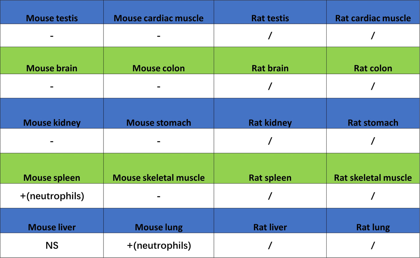
IHC shows positive staining in paraffin-embedded mouse spleen. Anti-CD177 antibody was used at 1/500 dilution, followed by a HRP Polymer for Rabbit IgG (ready to use). Counterstained with hematoxylin. Heat mediated antigen retrieval with Tris/EDTA buffer pH9.0 was performed before commencing with IHC staining protocol.
IHC shows positive staining in paraffin-embedded mouse lung. Anti-CD177 antibody was used at 1/500 dilution, followed by a HRP Polymer for Rabbit IgG (ready to use). Counterstained with hematoxylin. Heat mediated antigen retrieval with Tris/EDTA buffer pH9.0 was performed before commencing with IHC staining protocol.