WB result of CD11b Rabbit Polyclonal Antibody
Primary antibody: CD11b Rabbit Polyclonal Antibody at 1/1000 dilution
Lane 1: C2C12 whole cell lysate 20 μg
Lane 2: Raw 264.7 whole cell lysate 20 µg
Negative control: C2C12 whole cell lysate
Secondary antibody: Goat Anti-Rabbit IgG (H+L), HRP conjugated at 1/10000 dilution
Predicted MW: 127 kDa
Observed MW: 170 kDa
CD11b Rabbit Polyclonal Antibody
CD11b Rabbit Polyclonal Antibody
Price:
Regular price
$100 USD
Regular price
Sale price
$100 USD
Unit price
per
For shipping services or bulk orders, you may request a quotation.
Secure checkout with

View full details
Product Details
Product Details
Product Specification
| Host | Rabbit |
| Antigen | CD11b |
| Immunogen | Synthetic Peptide |
| Location | Cytoplasm |
| Accession | P05555 |
| Antibody Type | Polyclonal antibody |
| Isotype | IgG |
| Application | WB, IHC-P |
| Reactivity | Ms |
| Positive Sample | RAW264.7, mouse spleen, rat spleen |
| Purification | Immunogen Affinity |
| Concentration | 0.5 mg/ml |
| Conjugation | Unconjugated |
| Physical Appearance | Liquid |
| Storage Buffer | PBS, 40% Glycerol, 0.05% BSA, 0.02% sodium azide |
| Stability & Storage | 12 months from date of receipt / reconstitution, -20 °C as supplied |
Dilution
| application | dilution | species |
| WB | 1:1000 | Ms |
| IHC-P | 1:500 | Ms |
Background
CD11b (also known as the integrin alpha M subunit, ITGAM) is a type I transmembrane glycoprotein expressed primarily on the surface of monocytes, macrophages, neutrophils, NK cells, and some dendritic cells and brain microglia. Its extracellular domain non-covalently associates with the beta 2 integrin subunit (CD18) to form the heterodimeric integrin Mac-1 (also called complement receptor 3, CR3). As an adhesion molecule and signaling receptor, the CD11b/CD18 heterodimer recognizes a variety of ligands—including iC3b, ICAM-1, and fibrinogen—through the inserted (I) domain in the N-terminus of its alpha M subunit, thereby mediating leukocyte adhesion to vascular endothelium, transendothelial migration, and phagocytic clearance of complement-opsonized particles. Functionally, CD11b exhibits bidirectional signaling properties: on the one hand, it promotes phagocytosis and participates in inflammatory cell recruitment through recognition of iC3b; on the other hand, it can negatively regulate TLR and BCR signaling pathways, suppressing excessive inflammatory responses and maintaining immune tolerance. Clinical and genetic studies have shown that specific mutations in the ITGAM gene (such as R77H) can impair ligand binding and phagocytic function and are associated with susceptibility to systemic lupus erythematosus; in addition, the level of activated CD11b on the surface of neutrophils is positively correlated with disease activity in ANCA-associated vasculitis, suggesting its potential value as an inflammatory biomarker.
Picture
Picture
Western Blot
WB result of CD11b Rabbit Polyclonal Antibody
Primary antibody: CD11b Rabbit Polyclonal Antibody at 1/1000 dilution
Lane 1: mouse spleen lysate 20 μg
Secondary antibody: Goat Anti-Rabbit IgG (H+L), HRP conjugated at 1/10000 dilution
Predicted MW: 127 kDa
Observed MW: 170 kDa
WB result of CD11b Rabbit Polyclonal Antibody
Primary antibody: CD11b Rabbit Polyclonal Antibody at 1/1000 dilution
Lane 1: rat spleen lysate 20 μg
Secondary antibody: Goat Anti-Rabbit IgG (H+L), HRP conjugated at 1/10000 dilution
Predicted MW: 127 kDa
Observed MW: 170 kDa
Immunohistochemistry
IHC shows positive staining in paraffin-embedded mouse lung. Anti-CD11b antibody was used at 1/500 dilution, followed by a HRP Polymer for Rabbit IgG (ready to use). Counterstained with hematoxylin. Heat mediated antigen retrieval with Tris/EDTA buffer pH9.0 was performed before commencing with IHC staining protocol.
IHC shows positive staining in paraffin-embedded mouse spleen. Anti-CD11b antibody was used at 1/500 dilution, followed by a HRP Polymer for Rabbit IgG (ready to use). Counterstained with hematoxylin. Heat mediated antigen retrieval with Tris/EDTA buffer pH9.0 was performed before commencing with IHC staining protocol.
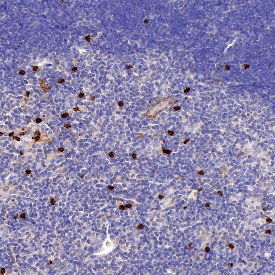
IHC shows positive staining in paraffin-embedded mouse thymus. Anti-CD11b antibody was used at 1/500 dilution, followed by a HRP Polymer for Rabbit IgG (ready to use). Counterstained with hematoxylin. Heat mediated antigen retrieval with Tris/EDTA buffer pH9.0 was performed before commencing with IHC staining protocol.