WB result of CCR9 Recombinant Rabbit mAb
Primary antibody: CCR9 Recombinant Rabbit mAb at 1/1000 dilution
Lane 1: unboiled HEK-293 whole cell lysate 20 μg
Lane 2: unboiled MOLT-4 whole cell lysate 20 μg
Low expression control: unboiled HEK-293 whole cell lysate
Secondary antibody: Goat Anti-Rabbit IgG (H+L), HRP conjugated at 1/10000 dilution
Predicted MW: 42 kDa
Observed MW: 50-60 kDa
This blot was developed with high sensitivity substrate
CCR9 Recombinant Rabbit mAb (S-2555-73)
CCR9 Recombinant Rabbit mAb (S-2555-73)
Price:
Regular price
$100 USD
Regular price
Sale price
$100 USD
Unit price
per
For shipping services or bulk orders, you may request a quotation.
Secure checkout with

View full details
Product Details
Product Details
Product Specification
| Host | Rabbit |
| Antigen | CD199 |
| Synonyms | C-C chemokine receptor type 9; C-C CKR-9; CC-CKR-9; CCR-9; G-protein coupled receptor 28; GPR-9-6; CDw199; GPR28; CCR9 |
| Immunogen | Recombinant Protein |
| Location | Cell membrane |
| Accession | P51686 |
| Clone Number | S-2555-73 |
| Antibody Type | Recombinant mAb |
| Isotype | IgG |
| Application | WB, FCM |
| Reactivity | Hu |
| Positive Sample | MOLT-4, Human PBMCs |
| Purification | Protein A |
| Concentration | 0.5 mg/ml |
| Conjugation | Unconjugated |
| Physical Appearance | Liquid |
| Storage Buffer | PBS, 40% Glycerol, 0.05% BSA, 0.02% sodium azide |
| Stability & Storage | 12 months from date of receipt / reconstitution, -20 °C as supplied |
Dilution
| application | dilution | species |
| WB | 1:1000 | Hu |
| FCM | 1:50 | Hu |
Background
CD199 (also designated CCR9) is a ~42 kDa seven-transmembrane G-protein-coupled chemokine receptor that binds the chemokine CCL25 (TECK), is strongly expressed in thymic cortex and small intestinal epithelium, and is highly expressed on CD4+CD8+ double-positive thymocytes and naive CD8+ T cells, as well as on small intestinal B cells, subsets of memory CD4+/CD8+ T cells, and dendritic cells, thereby directing T-cell development in the thymus and lymphocyte homing to gut-associated tissues via the CCR9/CCL25 axis, with dysregulation implicated in inflammatory bowel disease and celiac disease .
Picture
Picture
Western Blot
FC
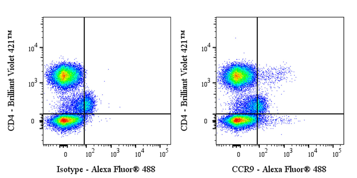
Flow cytometric analysis of human PBMCs (peripheral blood mononuclear cells) labeled with Anti-Human CCR9 antibody at 1/50 dilution (1 μg) / (right panel) compared with a Rabbit IgG Isotype Control / (left panel). Goat Anti- Rabbit IgG Alexa Fluor® 488 was used as the secondary antibody. Then cells were stained with CD4 - Brilliant Violet 421™ antibody separately. Flow cytometry and data analysis were performed using BD FACSymphony™ A1 and FlowJo™ software.