CA12 Rabbit Polyclonal Antibody
CA12 Rabbit Polyclonal Antibody
Price:
Regular price
$100 USD
Regular price
Sale price
$100 USD
Unit price
per
For shipping services or bulk orders, you may request a quotation.
Secure checkout with

View full details
Product Details
Product Details
Product Specification
| Host | Rabbit |
| Antigen | CA12 |
| Location | Cell membrane |
| Accession | O43570 |
| Antibody Type | Polyclonal antibody |
| Isotype | IgG |
| Application | WB, IHC-P, IF |
| Reactivity | Hu |
| Purification | Immunogen Affinity |
| Concentration | 2 mg/ml |
| Conjugation | Unconjugated |
| Physical Appearance | Liquid |
| Storage Buffer | PBS, 40% Glycerol, 0.05% BSA, 0.02% sodium azide |
| Stability & Storage | 12 months from date of receipt / reconstitution, -20 °C as supplied |
Dilution
| application | dilution | species |
| WB | 1:1000-1:2000 | Hu |
| IHC-P | 1:800 | Hu |
| IF | 1:800 | Hu |
Background
CA12 (carbonic anhydrase 12) is a zinc metalloenzyme that catalyzes the reversible hydration of carbon dioxide, participating in respiration, acid-base balance, and a variety of physiological processes. It belongs to the α-carbonic anhydrase family and is a single-pass type I membrane protein with an extracellular catalytic domain, existing as a homodimer. CA12 is highly expressed in normal kidney, colon, and pancreas, and its expression is regulated by hypoxia through the PHD/HIF-1α pathway. Functionally, CA12 plays a role in maintaining ion and pH homeostasis, and its deficiency can lead to isolated hyperchlorhidrosis. In the tumor microenvironment, CA12 is highly expressed on tumor-associated macrophages in hepatocellular carcinoma, where it sustains the survival and pro-metastatic functions of M2-like macrophages through HIF1α- and autocrine cytokine-dependent pathways; targeting CA12 can inhibit tumor progression and synergize with immune checkpoint blockade. In addition, CA12 exerts protective effects in nucleus pulposus cells, and its downregulation leads to reduced extracellular matrix synthesis and promotes intervertebral disc degeneration.
Picture
Picture
Western Blot
WB result of CA12 Rabbit Polyclonal Antibody
Primary antibody: CA12 Rabbit Polyclonal Antibody at 1/1000 dilution
Lane 1: untreated SK-OV-3 whole cell lysate 20 μg
Lane 2: untreated MCF7 whole cell lysate 20 μg
Lane 3: SK-OV-3 treated with PNGase F whole cell lysate 20 μg
Lane 4: MCF7 treated with PNGase F whole cell lysate 20 μg
Secondary antibody: Goat Anti-Rabbit IgG (H+L), HRP conjugated at 1/10000 dilution
Predicted MW: 39 kDa
Observed MW: 36, 40-50 kDa
Immunohistochemistry
IHC shows positive staining in paraffin-embedded human colon. Anti-CA12 antibody was used at 1/800 dilution, followed by a HRP Polymer for Rabbit IgG (ready to use). Counterstained with hematoxylin. Heat mediated antigen retrieval with Tris/EDTA buffer pH9.0 was performed before commencing with IHC staining protocol.
IHC shows positive staining in paraffin-embedded human kidney. Anti-CA12 antibody was used at 1/800 dilution, followed by a HRP Polymer for Rabbit IgG (ready to use). Counterstained with hematoxylin. Heat mediated antigen retrieval with Tris/EDTA buffer pH9.0 was performed before commencing with IHC staining protocol.
IHC shows positive staining in paraffin-embedded human endometrial cancer. Anti-CA12 antibody was used at 1/800 dilution, followed by a HRP Polymer for Rabbit IgG (ready to use). Counterstained with hematoxylin. Heat mediated antigen retrieval with Tris/EDTA buffer pH9.0 was performed before commencing with IHC staining protocol.
IHC shows positive staining in paraffin-embedded human renal cell carcinoma. Anti-CA12 antibody was used at 1/800 dilution, followed by a HRP Polymer for Rabbit IgG (ready to use). Counterstained with hematoxylin. Heat mediated antigen retrieval with Tris/EDTA buffer pH9.0 was performed before commencing with IHC staining protocol.
Immunofluorescence
IF shows positive staining in paraffin-embedded human kidney. Anti-CA12 antibody was used at 1/800 dilution (Green) and incubated overnight at 4°C. Goat polyclonal Antibody to Rabbit IgG - H&L (Alexa Fluor® 488) was used as secondary antibody at 1/1000 dilution. Counterstained with DAPI (Blue). Heat mediated antigen retrieval with EDTA buffer pH9.0 was performed before commencing with IF staining protocol.
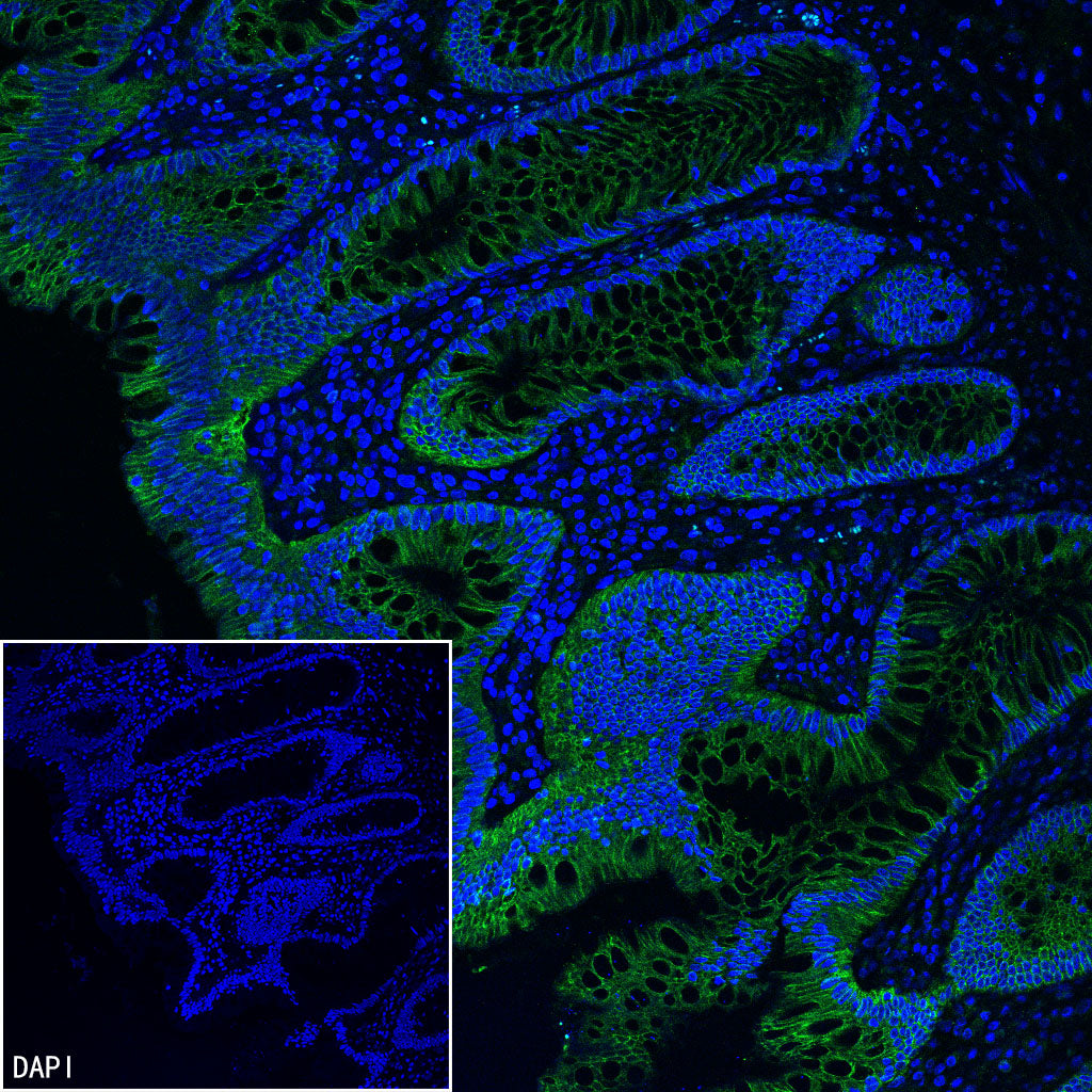
IF shows positive staining in paraffin-embedded human colon. Anti-CA12 antibody was used at 1/800 dilution (Green) and incubated overnight at 4°C. Goat polyclonal Antibody to Rabbit IgG - H&L (Alexa Fluor® 488) was used as secondary antibody at 1/1000 dilution. Counterstained with DAPI (Blue). Heat mediated antigen retrieval with EDTA buffer pH9.0 was performed before commencing with IF staining protocol.