WB result of BMI-1 Recombinant Rabbit mAb
Primary antibody: BMI-1 Recombinant Rabbit mAb at 1/1000 dilution
Lane 1: HeLa whole cell lysate 20 µg
Lane 2: A549 whole cell lysate 20 µg
Secondary antibody: Goat Anti-rabbit IgG, (H+L), HRP conjugated at 1/10000 dilution
Predicted MW: 37 kDa
Observed MW: 39 kDa
BMI-1 Recombinant Rabbit mAb (S-1075-252)
BMI-1 Recombinant Rabbit mAb (S-1075-252)
Price:
Regular price
$100 USD
Regular price
Sale price
$100 USD
Unit price
per
For shipping services or bulk orders, you may request a quotation.
Secure checkout with

View full details
Product Details
Product Details
Product Specification
| Host | Rabbit |
| Antigen | BMI-1 |
| Synonyms | Polycomb complex protein BMI-1, Polycomb group RING finger protein 4, RING finger protein 51, PCGF4, RNF51 |
| Immunogen | Synthetic Peptide |
| Location | Cytoplasm, Nucleus |
| Accession | P35226 |
| Clone Number | S-1075-252 |
| Antibody Type | Recombinant mAb |
| Isotype | IgG |
| Application | WB, ICC, ICFCM, ChIP |
| Reactivity | Hu, Ms, Rt |
| Predicted Reactivity | Bv, Ct, Ys |
| Purification | Protein A |
| Concentration | 0.5 mg/ml |
| Conjugation | Unconjugated |
| Physical Appearance | Liquid |
| Storage Buffer | PBS, 40% Glycerol, 0.05% BSA, 0.03% Proclin 300 |
| Stability & Storage | 12 months from date of receipt / reconstitution, -20 °C as supplied |
Dilution
| application | dilution | species |
| WB | 1:1000 | Hu, Ms, Rt |
| ICC | 1:500 | Hu, Ms |
| ICFCM | 1:50 | Hu, Ms |
| ChIP | 1:20-1:50 | Hu |
Background
Polycomb complex protein BMI-1, also known as B-cell specific Moloney murine leukemia virus integration site 1, is a component of the Polycomb repressive complex 1 (PRC1) that plays a significant role in gene silencing by regulating chromatin structure. BMI-1 is crucial for the self-renewal of both normal and cancer stem cells and is associated with the regulation of cell proliferation and senescence through the ink4a locus. It has been identified as an oncogene that can induce B- and T-cell leukemias and is frequently overexpressed in various types of cancer, including hematologic and solid cancers, suggesting its potential as a therapeutic target. BMI-1's involvement in cancer is not limited to its role in cell proliferation. It has also been shown to promote invasion and metastasis of cancer stem cells, particularly in pancreatic cancer, by activating the PI3K/AKT signaling pathway.
Picture
Picture
Western Blot
WB result of BMI-1 Recombinant Rabbit mAb
Primary antibody: BMI-1 Recombinant Rabbit mAb at 1/1000 dilution
Lane 1: NIH/3T3 whole cell lysate 20 µg
Secondary antibody: Goat Anti-rabbit IgG, (H+L), HRP conjugated at 1/10000 dilution
Predicted MW: 37 kDa
Observed MW: 39 kDa
WB result of BMI-1 Recombinant Rabbit mAb
Primary antibody: BMI-1 Recombinant Rabbit mAb at 1/1000 dilution
Lane 1: PC-12 whole cell lysate 20 µg
Secondary antibody: Goat Anti-rabbit IgG, (H+L), HRP conjugated at 1/10000 dilution
Predicted MW: 37 kDa
Observed MW: 40 kDa
FC
Flow cytometric analysis of 4% PFA fixed 90% methanol permeabilized HeLa (Human cervix adenocarcinoma epithelial cell) labelling BMI-1 antibody at 1/50 dilution (1 μg)/ (Red) compared with a Rabbit monoclonal IgG (Black) isotype control and an unlabelled control (cells without incubation with primary antibody and secondary antibody) (Blue). Goat Anti - Rabbit IgG Alexa Fluor® 488 was used as the secondary antibody.
Flow cytometric analysis of 4% PFA fixed 90% methanol permeabilized NIH/3T3 (Mouse embryonic fibroblast) labelling BMI-1 antibody at 1/50 dilution (1 μg)/ (Red) compared with a Rabbit monoclonal IgG (Black) isotype control and an unlabelled control (cells without incubation with primary antibody and secondary antibody) (Blue). Goat Anti - Rabbit IgG Alexa Fluor® 488 was used as the secondary antibody.
Immunocytochemistry
ICC shows positive staining in HeLa cells. Anti-BMI-1 antibody was used at 1/500 dilution (Green) and incubated overnight at 4°C. Goat polyclonal Antibody to Rabbit IgG - H&L (Alexa Fluor® 488) was used as secondary antibody at 1/1000 dilution. The cells were fixed with 100% ice-cold methanol and permeabilized with 0.1% PBS-Triton X-100. Nuclei were counterstained with DAPI (Blue). Counterstain with tubulin (Red).
ICC shows positive staining in NIH/3T3 cells. Anti-BMI-1 antibody was used at 1/500 dilution (Green) and incubated overnight at 4°C. Goat polyclonal Antibody to Rabbit IgG - H&L (Alexa Fluor® 488) was used as secondary antibody at 1/1000 dilution. The cells were fixed with 4% PFA and permeabilized with 0.1% PBS-Triton X-100. Nuclei were counterstained with DAPI (Blue). Counterstain with tubulin (Red).
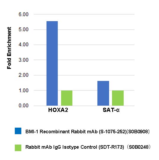
ChIP
Chromatin immunoprecipitation (ChIP) was performed on HeLa cells cross -
linked with 1% formaldehyde for 10 min, then chromatin was fragmented by sonication. Parallel reactions used BMI-1 Recombinant Rabbit mAb (S-1075-252) and Rabbit mAb IgG Isotype Control (SDT-R173) at 1:50 for immunoprecipitation.
Post - immunoprecipitation, both samples were washed, eluted, and cross - links reversed. Purified DNA was analyzed by qPCR.
qPCR showed the enrichment of HOXA2 and SAT-α in BMI-1 Recombinant
Rabbit mAb (S-1075-252)-immunoprecipitated sample.