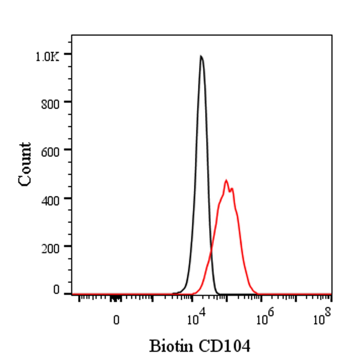
Flow cytometric analysis of mouse CD104 expression on bEnd.3 (Mouse brain endothelioma). bEnd.3 were stained with Biotin Rat IgG2a, κ Isotype Control (black line histogram) or SDT Biotin Rat Anti-Mouse CD104 Antibody (red line histogram) at 2 μl/test followed by Sav-iFluor 488. Flow cytometry and data analysis were performed using BD FACSymphony™ A1 and FlowJo™ software.
Biotin Rat Anti-Mouse CD104 Antibody (S-R716)
Biotin Rat Anti-Mouse CD104 Antibody (S-R716)
Price:
Regular price
$85 USD
Regular price
Sale price
$85 USD
Unit price
per
For shipping services or bulk orders, you may request a quotation.
Secure checkout with

View full details
Product Details
Product Details
Product Specification
| Host | Rat |
| Antigen | CD104 |
| Synonyms | Integrin beta-4; Itgb4 |
| Location | Cell membrane |
| Accession | A2A863 |
| Clone Number | S-R716 |
| Antibody Type | Rat mAb |
| Isotype | IgG2a,k |
| Application | FCM |
| Reactivity | Ms |
| Positive Sample | bEnd.3 |
| Purification | Protein G |
| Concentration | 0.5 mg/ml |
| Conjugation | Biotin |
| Physical Appearance | Liquid |
| Storage Buffer | PBS pH7.4, 0.09% sodium azide |
| Stability & Storage | 12 months from date of receipt / reconstitution, 2 to 8 °C as supplied |
Dilution
| application | dilution | species |
| FCM | 1μg per million cells in 100μl volume | Ms |
Background
CD104 is a key subunit of the integrin family, primarily functioning as a receptor for laminins and often associating with the alpha 6 subunit to form the α6β4 integrin complex. This transmembrane glycoprotein receptor plays a crucial role in mediating cell-matrix adhesion and signaling, which regulates gene expression, cell growth, and migration. CD104 is particularly important in maintaining the stability of hemidesmosomes, which anchor epithelial cells to the basement membrane. Additionally, its expression and activity are implicated in various biological processes, including wound healing, tissue integrity, and cancer progression. Abnormal mutations or dysregulation of CD104 have been associated with diseases such as epidermolysis bullosa with pyloric atresia.
Picture
Picture
FC