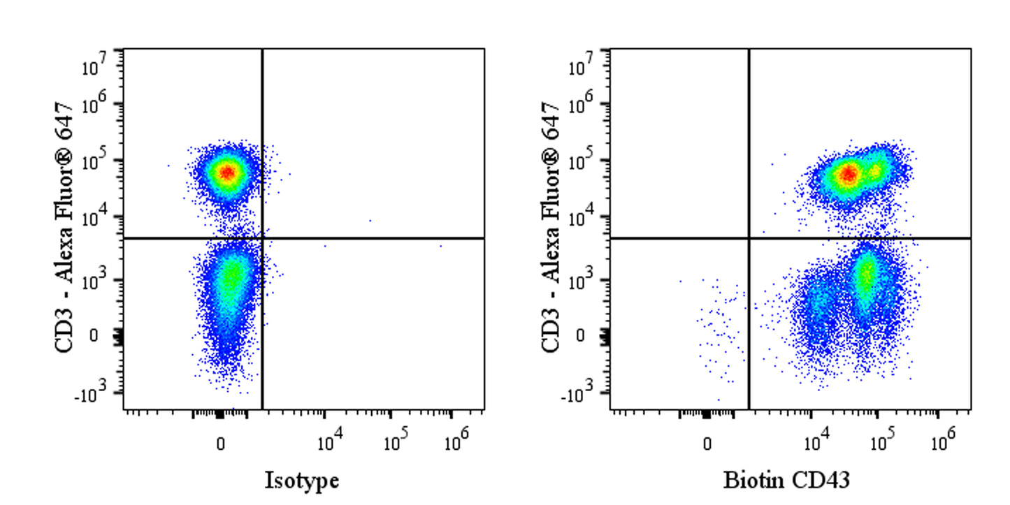
Flow cytometric analysis of human peripheral blood leukocytes fixed with 4% paraformaldehyde and permeabilized with 0.1% Tween. Cells were stained with Alexa Fluor® 647 Mouse anti-human CD3 Antibody (right panel) and either Biotin Rabbit IgG Isotype Control (left panel) or Biotin Rabbit Anti-Human CD43 Antibody (right panel) at 5 μl/test followed by Sav-iFluor 488. Total viable cells, as determined by Fixable Viability Dye 452 (S0B88801), were used for analysis. Flow cytometry and data analysis were performed using BD FACSymphony™ A1 and FlowJo™ software.
Biotin Rabbit Anti-Human CD43 Antibody (S-R101)
Biotin Rabbit Anti-Human CD43 Antibody (S-R101)
Price:
Regular price
$70 USD
Regular price
Sale price
$70 USD
Unit price
per
For shipping services or bulk orders, you may request a quotation.
Secure checkout with

View full details
Product Details
Product Details
Product Specification
| Host | Rabbit |
| Antigen | CD43 |
| Synonyms | Leukosialin; GPL115; Galactoglycoprotein (GALGP); Leukocyte sialoglycoprotein; Sialophorin; SPN |
| Location | Membrane |
| Accession | P16150 |
| Clone Number | S-R101 |
| Antibody Type | Recombinant mAb |
| Isotype | IgG |
| Isotype Control | S0B8273 |
| Application | ICFCM |
| Reactivity | Hu |
| Positive Sample | human peripheral blood leukocytes |
| Purification | Protein A |
| Concentration | 0.2 mg/ml |
| Conjugation | Biotin |
| Physical Appearance | Liquid |
| Storage Buffer | PBS pH7.4, 0.09% sodium azide |
| Stability & Storage | 12 months from date of receipt / reconstitution, 2 to 8 °C as supplied |
Dilution
| application | dilution | species |
| ICFCM | 5μl per million cells in 100μl volume | Hu |
Background
CD43, also known as Leukosialin or Sialophorin, is a type I transmembrane glycoprotein that is highly expressed on the surface of leukocytes (such as T cells, monocytes, and granulocytes) but is absent on resting naive B cells and erythrocytes. Structurally, it features a unique, negatively charged, rigid rod-like extracellular domain heavily modified by approximately 80 O-glycans, along with a cytoplasmic tail containing potential phosphorylation sites that can interact with cytoskeletal linker proteins (such as ERM family members). Functionally, CD43 plays a dual role as both an "anti-adhesion" molecule and a signaling regulator: its bulky extracellular domain serves as a physical barrier that negatively regulates integrin- and L-selectin-mediated cell adhesion and homing through steric hindrance, while its cytoplasmic domain transmits signals to finely regulate T cell activation, proliferation, apoptosis, and the contraction of immune responses. Furthermore, dysregulation of CD43 is closely associated with disease—it is defective or absent in patients with Wiskott-Aldrich syndrome. In various cancer cells, such as acute myeloid leukemia, highly expressed CD43 and its surface sialylated glycosylation modifications form a "glyco-immune barrier" that physically impedes contact between immune effector cells (including macrophages and NK cells) and tumor cells, thereby helping cancer cells achieve immune evasion. Given its central role in immune regulation and tumor immune escape, CD43 has emerged as a highly promising novel target for cancer immunotherapy.
Picture
Picture
FC