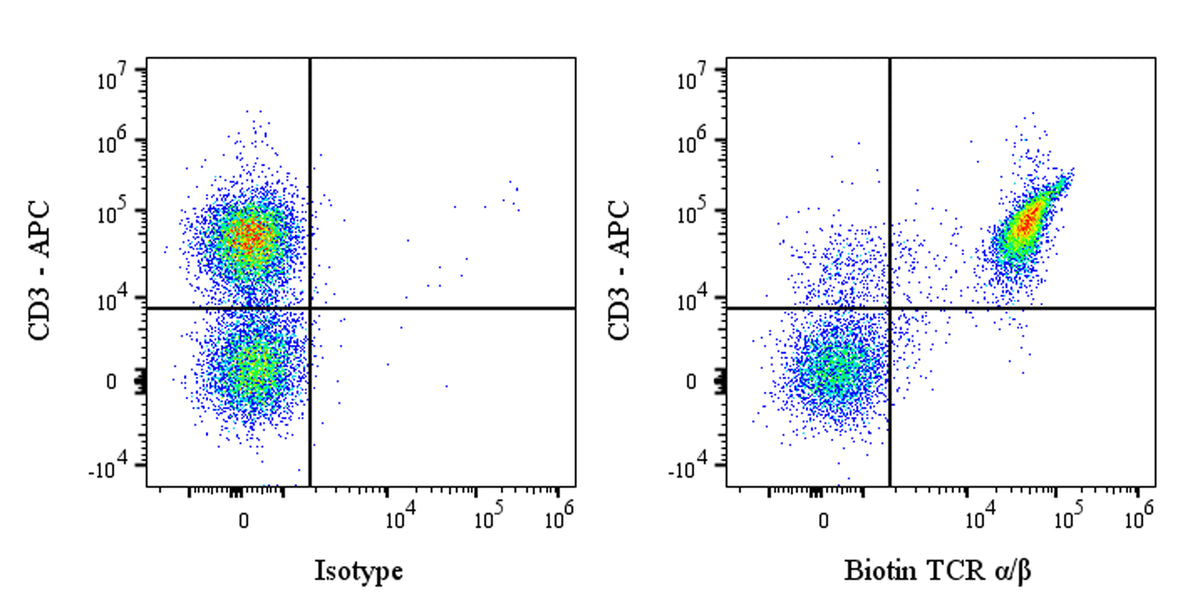
Flow cytometric analysis of TCR α/β expression on SD rat splenocytes. SD rat splenocytes were stained with APC Mouse Anti-Rat CD3 Antibody and either Biotin Mouse IgG1, k Isotype Control (left panel) or SDT Biotin Mouse Anti-Rat TCR α/β Antibody (right panel) at 2 μl/test followed by Sav-iFluor 488. Flow cytometry and data analysis were performed using Agilent NovoCyte Quanteon and FlowJo™ software.
Biotin Mouse Anti-Rat TCR α/β Antibody (R73)
Biotin Mouse Anti-Rat TCR α/β Antibody (R73)
Price:
Regular price
$130 USD
Regular price
Sale price
$130 USD
Unit price
per
For shipping services or bulk orders, you may request a quotation.
Secure checkout with

View full details
Product Details
Product Details
Product Specification
| Host | Mouse |
| Antigen | TCR α/β |
| Location | Cell membrane |
| Accession | A0A8I6ABG9 |
| Clone Number | R73 |
| Antibody Type | Mouse mAb |
| Isotype | IgG1,k |
| Isotype Control | S0B5431 |
| Application | FCM |
| Reactivity | Rt |
| Positive Sample | SD rat splenocytes |
| Purification | Protein G |
| Concentration | 0.5 mg/ml |
| Conjugation | Biotin |
| Physical Appearance | Liquid |
| Storage Buffer | PBS pH7.4, 0.09% sodium azide |
| Stability & Storage | 12 months from date of receipt / reconstitution, 2 to 8 °C as supplied |
Dilution
| application | dilution | species |
| FCM | 1μg per million cells in 100μl volume | Rt |
Background
TCR α/β protein, the defining heterodimer of the αβ T-cell lineage, is a disulfide-linked transmembrane receptor whose extracellular amino-terminal variable (Vα/Vβ) and constant (Cα/Cβ) Ig-like domains collectively form an antigen-binding groove that recognizes a composite ligand consisting of a peptide non-covalently nestled within the groove of an MHC molecule; this interaction is stabilized by two paired immunoglobulin superfamily domains and anchored to the T-cell membrane via stalk, transmembrane and short cytoplasmic regions that associate with CD3 γε, δε and ζζ signaling subunits, so that ligand-induced conformational change in the αβ heterodimer triggers phosphorylation of ITAMs and initiates the calcium- and kinase-dependent cascade leading to T-cell activation, proliferation and differentiation, thereby making the TCR α/β protein the central sensor that converts extracellular peptide-MHC information into intracellular immune decisions.
Picture
Picture
FC