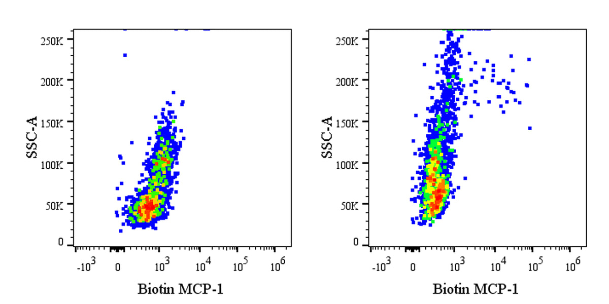
Flow cytometric analysis of MCP-1/CCL2 expression in stimulated Human PBMCs (human peripheral blood mononuclear cells). Human PBMCs were cultured 6hr with 10ng/ml lipopolysaccharide in the presence of 1 μg/ml BFA (right panel) or untreated (left panel). The PBMCs were harvested and fixed with 4% PFA and permeabilized with Intracellular Fixation & Permeabilization Buffer Set. The cells were then stained with Biotin Mouse Anti-Human MCP-1/CCL2 Antibody at 5 μl/test followed by Sav-PE. Flow cytometry and data analysis were performed using BD FACSymphony™ A1 and FlowJo™ software.
Biotin Mouse Anti-Human MCP-1/CCL2 Antibody (5D3-F7)
Biotin Mouse Anti-Human MCP-1/CCL2 Antibody (5D3-F7)
Price:
Regular price
$180 USD
Regular price
Sale price
$180 USD
Unit price
per
For shipping services or bulk orders, you may request a quotation.
Secure checkout with

View full details
Product Details
Product Details
Product Specification
| Host | Mouse |
| Antigen | CCL2 |
| Location | Secreted |
| Accession | P13500 |
| Clone Number | 5D3-F7 |
| Antibody Type | Mouse mAb |
| Isotype Control | S0B5431 |
| Application | ICFCM |
| Reactivity | Hu |
| Positive Sample | Human PBMCs stimulated with LPS and BFA |
| Purification | Protein A |
| Concentration | 0.2 mg/ml |
| Conjugation | Biotin |
| Physical Appearance | Liquid |
| Storage Buffer | PBS pH7.4, 0.09% sodium azide |
| Stability & Storage | 12 months from date of receipt / reconstitution, 2 to 8 °C as supplied |
Dilution
| application | dilution | species |
| ICFCM | 5μl per million cells in 100μl volume | Hu |
Background
MCP-1, or Monocyte Chemoattractant Protein-1 (also known as CCL2), is a key member of the CC chemokine subfamily. It is primarily secreted by various cell types, including monocytes and endothelial cells. Its core function is to potently attract and activate monocytes by binding to its receptor CCR2, guiding them to cross the vascular endothelium and migrate to sites of inflammation or injury. While MCP-1 is indispensable for host defense and immune surveillance, its overexpression or aberrant expression drives the pathology of numerous diseases. For instance, in osteoarthritis, it promotes the polarization of monocytes toward pro-inflammatory M1-type macrophages; it also plays significant roles in atherosclerosis, tumor microenvironment remodeling, and autoimmune diseases such as systemic lupus erythematosus. Furthermore, recent studies have uncovered non-chemotactic functions of MCP-1 in the nervous system, where it can enhance excitatory synaptic transmission in neurons through specific signaling pathways, indicating its complex involvement in neuroinflammation and pain modulation.
Picture
Picture
FC