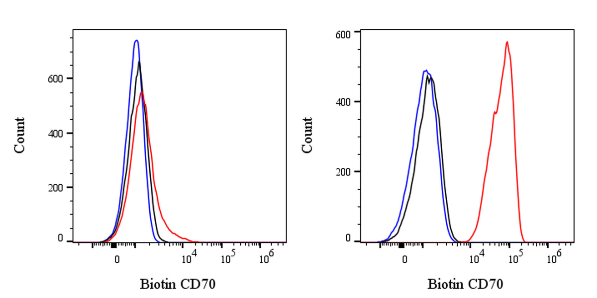
Flow cytometric analysis of Human CD70 expression on U266B1 cells. Cells from the U266B1 (Human multiple myeloma B lymphocyte, right) or Jurkat (Human T cell leukemia T lymphocyte, left) were stained with either Biotin Mouse IgG3, κ Isotype Control (black line histogram) or SDT Biotin Mouse Anti-Human CD70 Antibody (red line histogram) at 5 μl/test followed by Sav-iFluor 488, cells without incubation with primary antibody and secondary antibody (blue line histogram) were used as unlabeled control. Flow cytometry and data analysis were performed using Agilent NovoCyte Quanteon and FlowJo™ software.
Biotin Mouse Anti-Human CD70 Antibody (Ki-24)
Biotin Mouse Anti-Human CD70 Antibody (Ki-24)
Price:
Regular price
$170 USD
Regular price
Sale price
$170 USD
Unit price
per
For shipping services or bulk orders, you may request a quotation.
Secure checkout with

View full details
Product Details
Product Details
Product Specification
| Host | Mouse |
| Antigen | CD70 |
| Location | Cell membrane |
| Accession | P32970 |
| Clone Number | Ki-24 |
| Antibody Type | Mouse mAb |
| Application | FCM |
| Reactivity | Hu |
| Positive Sample | U266B1 |
| Purification | Protein A |
| Concentration | 0.2 mg/ml |
| Conjugation | Biotin |
| Physical Appearance | Liquid |
| Storage Buffer | PBS pH7.4, 0.09% sodium azide |
| Stability & Storage | 12 months from date of receipt / reconstitution, 2 to 8 °C as supplied |
Dilution
| application | dilution | species |
| FCM | 5μl per million cells in 100μl volume | Hu |
Background
CD70, also known as CD27 ligand, is a type Ⅱ transmembrane protein that plays a crucial role in the immune system. It is primarily expressed on antigen - presenting cells (APCs) such as dendritic cells, macrophages, and B cells, as well as on activated T cells. CD70 interacts with its receptor CD27, which is mainly found on T cells, B cells, and natural killer (NK) cells. This interaction provides important co - stimulatory signals that promote T cell activation, proliferation, and differentiation, and also enhances B cell and NK cell functions. Additionally, CD70 is involved in the regulation of immune responses, including the maintenance of immune homeostasis and the induction of immune - mediated tumor cell killing. Abnormal expression of CD70 has been associated with various diseases, such as autoimmune disorders and certain types of cancers. Therefore, CD70 is considered a promising target for the development of immunotherapies.
Picture
Picture
FC