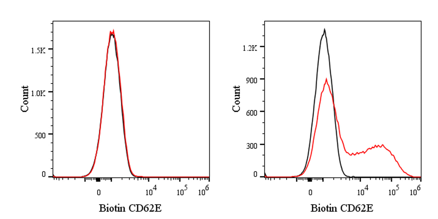
Flow cytometric analysis of CD62E expression on HUVEC (Human umbilical vein endothelial cells) (left panel) or TNF-α activated HUVEC (right panel). HUVEC treated 6 hours with 25 ng/ml TNF-α were stained with Biotin Mouse IgG1, k Isotype Control (black line histogram) and SDT Biotin Mouse Anti-Human CD62E Antibody (red line histogram) at 5 μg/test followed by Sav-APC. Flow cytometry and data analysis were performed using BD FACSymphony™ A1 and FlowJo™ software.
Biotin Mouse Anti-Human CD62E Antibody (S-2909)
Biotin Mouse Anti-Human CD62E Antibody (S-2909)
Price:
Regular price
$135 USD
Regular price
Sale price
$135 USD
Unit price
per
For shipping services or bulk orders, you may request a quotation.
Secure checkout with

View full details
Product Details
Product Details
Product Specification
| Host | Mouse |
| Antigen | CD62E |
| Synonyms | E-selectin; CD62 antigen-like family member E; Endothelial leukocyte adhesion molecule 1 (ELAM-1); Leukocyte-endothelial cell adhesion molecule 2 (LECAM2); ELAM1; SELE |
| Location | Cell membrane |
| Accession | P16581 |
| Clone Number | S-2909 |
| Antibody Type | Mouse mAb |
| Isotype | IgG1,k |
| Application | FCM |
| Reactivity | Hu |
| Positive Sample | TNF-α activated HUVEC |
| Purification | Protein G |
| Concentration | 0.2 mg/ml |
| Conjugation | Biotin |
| Physical Appearance | Liquid |
| Storage Buffer | PBS pH7.4, 0.03% Proclin 300 |
| Stability & Storage | 12 months from date of receipt / reconstitution, 2 to 8 °C as supplied |
Dilution
| application | dilution | species |
| FCM | 5μl per million cells in 100μl volume | Hu |
Background
CD62E, also known as E-selectin or endothelial-leukocyte adhesion molecule-1 (ELAM-1), is a ~115 kDa calcium-dependent type-I transmembrane glycoprotein of the selectin family expressed exclusively on cytokine-activated vascular endothelium where it mediates the first tethering and rolling of circulating leukocytes by binding sialyl-Lewis X-decorated ligands such as PSGL-1 (SELPLG) on neutrophils, monocytes, and effector/memory T cells, thereby orchestrating extravasation during acute and chronic inflammation; the molecule comprises an N-terminal C-type lectin domain, an epidermal growth factor-like motif, six complement-binding (sushi/CCP) repeats, a single transmembrane segment, and a short cytoplasmic tail, is transcriptionally up-regulated within 2–4 h of stimulation by IL-1β, TNF-α, or LPS via an NF-κB–dependent pathway, and also participates in angiogenesis, tumor-cell metastasis, and vascular repair, making it both a biomarker and therapeutic target in cardiovascular disease, sepsis, and cancer.
Picture
Picture
FC