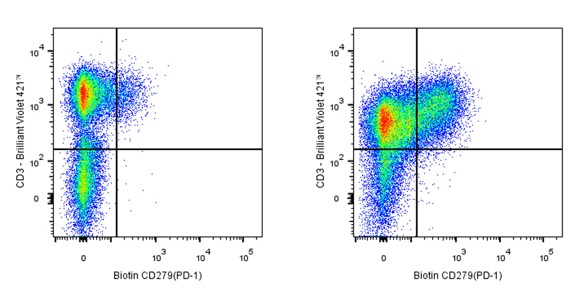
Flow cytometric analysis of human peripheral blood lymphocytes were stained with stimulated 2 days with 5 μg/ml PHA (Right panel) or unstimulated (Left panel) was stained with Alexa Fluor® 488 Mouse Anti-Human CD3 and SDT Biotin Mouse Anti-Human CD279 (PD-1) Antibody at 5 μl/test followed by Sav-iFluor 488. Flow cytometry and data analysis were performed using BD FACSymphony™ A1 and FlowJo™ software.
Biotin Mouse Anti-Human CD279 (PD-1) Antibody (EH12.2H7)
Biotin Mouse Anti-Human CD279 (PD-1) Antibody (EH12.2H7)
Price:
Regular price
$197 USD
Regular price
Sale price
$197 USD
Unit price
per
For shipping services or bulk orders, you may request a quotation.
Secure checkout with

View full details
Product Details
Product Details
Product Specification
| Host | Mouse |
| Synonyms | Programmed cell death protein 1; Protein PD-1; hPD-1; PD1; PDCD1 |
| Location | Cell membrane |
| Accession | Q15116 |
| Clone Number | EH12.2H7 |
| Antibody Type | Mouse mAb |
| Isotype | IgG1,k |
| Application | FCM |
| Reactivity | Hu |
| Purification | Protein G |
| Concentration | 0.2 mg/ml |
| Conjugation | Biotin |
| Physical Appearance | Liquid |
| Storage Buffer | PBS pH7.4, 0.03% Proclin 300 |
| Stability & Storage | 12 months from date of receipt / reconstitution, 2 to 8 °C as supplied |
Dilution
| application | dilution | species |
| FCM | 5μl per million cells in 100μl volume | Hu |
Background
Programmed cell death protein 1 (PD-1), also known as CD279, is a type I transmembrane protein belonging to the CD28 immunoglobulin superfamily. It is encoded by the Pdcd1 gene and consists of 288 amino acids, including an extracellular IgV-like domain, a transmembrane region, and a cytoplasmic tail. PD-1 is expressed on activated T cells, B cells, macrophages, dendritic cells, and natural killer cells. Its cytoplasmic domain contains two tyrosine residues that form an immunoreceptor tyrosine-based inhibitory motif (ITIM) and an immunoreceptor tyrosine-based switch motif (ITSM), which play key roles in inhibiting T cell receptor signaling. PD-1 interacts with its ligands PD-L1 and PD-L2 to negatively regulate immune responses, contributing to peripheral tolerance and preventing autoimmune diseases. However, this inhibitory function can also suppress anti-tumor immunity, making PD-1 a promising target for cancer immunotherapy.
Picture
Picture
FC