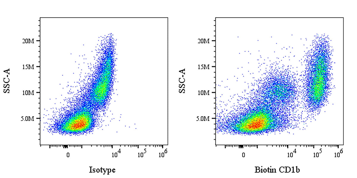
Flow cytometric analysis of CD1b expression on human PBMCs (human peripheral blood mononuclear cells) spiked with Molt-4 (human lymphoblastic leukemia T lymphoblast). The cells were stained with either Biotin Mouse IgG1, κ Isotype Control (left panel) or SDT Biotin Mouse Anti-Human CD1b Antibody (right panel) at 5 μl/test followed by Sav-iFluor 488. Flow cytometry and data analysis were performed using Agilent NovoCyte Quanteon and FlowJo™ software.
Biotin Mouse Anti-Human CD1b Antibody (SN13)
Biotin Mouse Anti-Human CD1b Antibody (SN13)
Price:
Regular price
$85 USD
Regular price
Sale price
$85 USD
Unit price
per
For shipping services or bulk orders, you may request a quotation.
Secure checkout with

View full details
Product Details
Product Details
Product Specification
| Host | Mouse |
| Antigen | CD1b |
| Synonyms | T-cell surface glycoprotein CD1b; CD1B |
| Location | Cell membrane |
| Accession | P29016 |
| Clone Number | SN13 |
| Antibody Type | Mouse mAb |
| Isotype | IgG1,k |
| Isotype Control | S0B5431 |
| Application | FCM |
| Reactivity | Hu |
| Positive Sample | Human PBMCs spiked with Molt-4 cells |
| Purification | Protein G |
| Concentration | 0.2 mg/ml |
| Conjugation | Biotin |
| Physical Appearance | Liquid |
| Storage Buffer | PBS pH7.4, 0.09% sodium azide |
| Stability & Storage | 12 months from date of receipt / reconstitution, 2 to 8 °C as supplied |
Dilution
| application | dilution | species |
| FCM | 5μl per million cells in 100μl volume | Hu |
Background
CD1b is a glycoprotein that plays a critical sentinel role in the human immune system. It belongs to the CD1 family and functions similarly to classical MHC class I molecules, but with a fundamental difference: instead of presenting protein fragments (peptides), CD1b is specialized in binding and presenting lipid and glycolipid antigens to T cells. The antigen-binding groove of CD1b has a unique structure, composed of four interconnected hydrophobic pockets (A', C', F', and T') that are relatively large in volume, allowing it to accommodate structurally diverse lipids, including molecules with significant variations in carbon chain length (ranging from C12 to C80)—such as mycolic acid, glucose monomycolate (GMM), and phosphatidylinositol mannoside (PIM) found in the cell walls of mycobacteria. To efficiently load long-chain lipid antigens like those derived from Mycobacterium tuberculosis, CD1b relies on signaling motifs in its cytoplasmic tail to internalize from the cell surface and traffic to acidic endosomal/lysosomal compartments, where lipid loading is facilitated by the acidic environment and accessory proteins, after which it returns to the cell surface to present antigens to T cells. Consequently, CD1b plays an indispensable role in the immune defense against mycobacterial infections such as tuberculosis, with its expression and function dynamically modulated during infection to mobilize T-cell-mediated specific killing responses against lipid antigens.
Picture
Picture
FC