WB result of Bcl 7A Recombinant Rabbit mAb
Primary antibody: Bcl 7A Recombinant Rabbit mAb at 1/1000 dilution
Lane 1: Daudi whole cell lysate 20 μg
Lane 2: HEK-293 whole cell lysate 20 μg
Lane 3: HeLa whole cell lysate 20 μg
Lane 4: HDLM-2 whole cell lysate 20 μg
Secondary antibody: Goat Anti-Rabbit IgG (H+L), HRP conjugated at 1/10000 dilution
Predicted MW: 22 kDa
Observed MW: 38, 40 kDa
Bcl 7A Recombinant Rabbit mAb (S-6523)
Bcl 7A Recombinant Rabbit mAb (S-6523)
Price:
Regular price
$100 USD
Regular price
Sale price
$100 USD
Unit price
per
For shipping services or bulk orders, you may request a quotation.
Secure checkout with

View full details
Product Details
Product Details
Product Specification
| Host | Rabbit |
| Antigen | Bcl 7A |
| Synonyms | B-cell CLL/lymphoma 7 protein family member A; BCL7A |
| Location | Nucleus |
| Accession | Q4VC05 |
| Clone Number | S-6523 |
| Antibody Type | Recombinant mAb |
| Isotype | IgG |
| Application | WB, IHC-P, ICC |
| Reactivity | Hu, Ms, Rt |
| Positive Sample | Daudi, HEK-293, HeLa, HDLM-2, Neuro-2a, mouse brain, C6, rat brain |
| Purification | Protein A |
| Concentration | 0.5 mg/ml |
| Conjugation | Unconjugated |
| Physical Appearance | Liquid |
| Storage Buffer | PBS, 40% Glycerol, 0.05% BSA, 0.02% sodium azide |
| Stability & Storage | 12 months from date of receipt / reconstitution, -20 °C as supplied |
Dilution
| application | dilution | species |
| WB | 1:1000 | Hu, Ms, Rt |
| IHC-P | 1:2000 | Hu, Ms, Rt |
| ICC | 1:500 | Hu, Ms |
Background
BCL7A is a relatively recently identified subunit of the mammalian SWI/SNF chromatin remodeling complex, belonging to the BCL7 protein family (BCL7A/B/C). It is specifically present in multicellular animals but absent from the single-cell versions of the complex. It is a shared component of all three human SWI/SNF variants—cBAF, PBAF, and ncBAF—and its N-terminal double-stranded β-sheet associates with the ACTB-ACTL6A dimer, which in turn links to the HSA domain of BRG1, mediating its integration into the complex. Cryo-electron microscopy structural studies published in 2025 revealed that BCL7A, through an arginine anchoring motif in its N-terminal α-helix (rich in basic residues such as R4, R7, and R11), binds with high affinity to the acidic patch on the surface of the histone H2A-H2B of the nucleosome core particle. Functionally, the N-terminal α-helix of BCL7A plays a key regulatory role in cBAF-mediated nucleosome sliding and histone exchange; deletion of this region significantly impairs chromatin remodeling activity without affecting the basal ATPase activity of BRG1. At the pathological level, BCL7A is frequently mutated in various hematological malignancies (especially diffuse large B-cell lymphoma), and mutations in its N-terminal domain can impair its tumor-suppressive function; cancer-associated BCL7A mutations also weaken its ability to bind the acidic patch of the nucleosome.
Picture
Picture
Western Blot
WB result of Bcl 7A Recombinant Rabbit mAb
Primary antibody: Bcl 7A Recombinant Rabbit mAb at 1/1000 dilution
Lane 1: Neuro-2a whole cell lysate 20 μg
Lane 2: mouse brain lysate 20 μg
Secondary antibody: Goat Anti-Rabbit IgG (H+L), HRP conjugated at 1/10000 dilution
Predicted MW: 22 kDa
Observed MW: 38 kDa
WB result of Bcl 7A Recombinant Rabbit mAb
Primary antibody: Bcl 7A Recombinant Rabbit mAb at 1/1000 dilution
Lane 1: C6 whole cell lysate 20 μg
Lane 2: rat brain lysate 20 μg
Secondary antibody: Goat Anti-Rabbit IgG (H+L), HRP conjugated at 1/10000 dilution
Predicted MW: 22 kDa
Observed MW: 38 kDa
Immunohistochemistry
IHC shows positive staining in paraffin-embedded human spleen. Anti-Bcl7A antibody was used at 1/2000 dilution, followed by a HRP Polymer for Rabbit IgG (ready to use). Counterstained with hematoxylin. Heat mediated antigen retrieval with Tris/EDTA buffer pH9.0 was performed before commencing with IHC staining protocol.
IHC shows positive staining in paraffin-embedded human tonsil. Anti-Bcl7A antibody was used at 1/2000 dilution, followed by a HRP Polymer for Rabbit IgG (ready to use). Counterstained with hematoxylin. Heat mediated antigen retrieval with Tris/EDTA buffer pH9.0 was performed before commencing with IHC staining protocol.
IHC shows positive staining in paraffin-embedded human colon cancer. Anti-Bcl7A antibody was used at 1/2000 dilution, followed by a HRP Polymer for Rabbit IgG (ready to use). Counterstained with hematoxylin. Heat mediated antigen retrieval with Tris/EDTA buffer pH9.0 was performed before commencing with IHC staining protocol.
IHC shows positive staining in paraffin-embedded mouse brain. Anti-Bcl7A antibody was used at 1/2000 dilution, followed by a HRP Polymer for Rabbit IgG (ready to use). Counterstained with hematoxylin. Heat mediated antigen retrieval with Tris/EDTA buffer pH9.0 was performed before commencing with IHC staining protocol.
IHC shows positive staining in paraffin-embedded rat brain. Anti-Bcl7A antibody was used at 1/2000 dilution, followed by a HRP Polymer for Rabbit IgG (ready to use). Counterstained with hematoxylin. Heat mediated antigen retrieval with Tris/EDTA buffer pH9.0 was performed before commencing with IHC staining protocol.
Immunocytochemistry
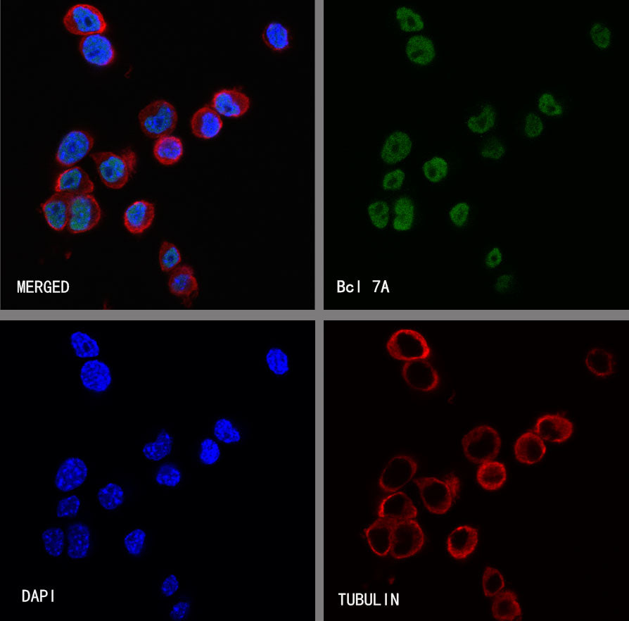
ICC shows positive staining in HEK293 cells. Anti-Bcl 7A antibody was used at 1/500 dilution (Green) and incubated overnight at 4°C. Goat polyclonal Antibody to Rabbit IgG - H&L (Alexa Fluor® 488) was used as secondary antibody at 1/1000 dilution. The cells were fixed with 4% PFA and permeabilized with 0.1% PBS-Triton X-100. Nuclei were counterstained with DAPI (Blue). Counterstain with tubulin (Red).
ICC shows positive staining in Neuro-2a cells. Anti-Bcl 7A antibody was used at 1/500 dilution (Green) and incubated overnight at 4°C. Goat polyclonal Antibody to Rabbit IgG - H&L (Alexa Fluor® 488) was used as secondary antibody at 1/1000 dilution. The cells were fixed with 4% PFA and permeabilized with 0.1% PBS-Triton X-100. Nuclei were counterstained with DAPI (Blue). Counterstain with tubulin (Red).