WB result of Bcl-2 Recombinant Rabbit mAb
Primary antibody: Bcl-2 Recombinant Rabbit mAb at 1/1000 dilution
Primary antibody incubation conditions: overnight at 4°C
Lane 1: Neuro-2a whole cell lysate 20 µg
Lane 2: CTLL-2 whole cell lysate 20 µg
Negative control: Neuro-2a whole cell lysate
Secondary antibody: Goat Anti-Rabbit IgG (H+L), HRP conjugated at 1/10000 dilution
Predicted MW: 26 kDa
Observed MW: 24 kDa
Bcl-2 Recombinant Rabbit mAb (S-3910-3)
Bcl-2 Recombinant Rabbit mAb (S-3910-3)
Price:
Regular price
$100 USD
Regular price
Sale price
$100 USD
Unit price
per
For shipping services or bulk orders, you may request a quotation.
Secure checkout with

View full details
Product Details
Product Details
Product Specification
| Host | Rabbit |
| Antigen | Bcl-2 |
| Immunogen | Synthetic Peptide |
| Location | Cytoplasm |
| Accession | P10417 |
| Clone Number | S-3910-3 |
| Antibody Type | Recombinant mAb |
| Isotype | IgG |
| Application | WB, IHC-P |
| Reactivity | Ms, Rt, Mk |
| Positive Sample | CTLL2, C2C12, NIH/3T3, mouse spleen, rat spleen, rat kidney, COS-7 |
| Purification | Protein A |
| Concentration | 0.5 mg/ml |
| Conjugation | Unconjugated |
| Physical Appearance | Liquid |
| Storage Buffer | PBS, 40% Glycerol, 0.05% BSA, 0.02% sodium azide |
| Stability & Storage | 12 months from date of receipt / reconstitution, -20 °C as supplied |
Dilution
| application | dilution | species |
| WB | 1:1000 | Ms, Rt |
| IHC-P | 1:200 | Ms, Rt |
Background
Bcl-2 (B-cell lymphoma/leukemia-2) is an anti-apoptotic protein localized to the outer mitochondrial membrane, endoplasmic reticulum, and nuclear envelope. It consists of 239 amino acids with a molecular weight of approximately 25–26 kD, and its structure contains four conserved homology domains (BH1 to BH4), a flexible loop region, and a C-terminal transmembrane anchoring domain. As the prototype member of the anti-apoptotic subfamily within the Bcl-2 protein family, the core function of Bcl-2 is to bind and inhibit the activity of pro-apoptotic proteins Bax and Bak through a hydrophobic groove formed by its BH1–BH3 domains, thereby blocking mitochondrial outer membrane permeabilization and the release of cytochrome c, and preventing caspase cascade activation and the execution of apoptosis. Bcl-2 was initially discovered because of overexpression caused by the t(14;18) chromosomal translocation in B-cell lymphoma. Its abnormal high expression enables tumor cells to evade programmed cell death, making it an important pathogenic mechanism in various hematological malignancies and solid tumors, and it is also closely associated with chemotherapy resistance and radiotherapy resistance. Currently, BH3 mimetics targeting Bcl-2 (such as venetoclax) have achieved significant clinical success in the treatment of chronic lymphocytic leukemia and acute myeloid leukemia, becoming a successful paradigm for cancer therapy by targeting the apoptotic pathway.
Picture
Picture
Western Blot
WB result of Bcl-2 Recombinant Rabbit mAb
Primary antibody: Bcl-2 Recombinant Rabbit mAb at 1/1000 dilution
Primary antibody incubation conditions: overnight at 4°C
Lane 1: NIH/3T3 whole cell lysate 20 µg
Lane 2: C2C12 whole cell lysate 20 µg
Lane 3: mouse spleen lysate 20 µg
Secondary antibody: Goat Anti-Rabbit IgG (H+L), HRP conjugated at 1/10000 dilution
Predicted MW: 26 kDa
Observed MW: 24 kDa
This blot was developed with high sensitivity substrate
WB result of Bcl-2 Recombinant Rabbit mAb
Primary antibody: Bcl-2 Recombinant Rabbit mAb at 1/1000 dilution
Primary antibody incubation conditions: overnight at 4°C
Lane 1: rat spleen lysate 20 µg
Lane 2: rat kidney lysate 20 µg
Secondary antibody: Goat Anti-Rabbit IgG (H+L), HRP conjugated at 1/10000 dilution
Predicted MW: 26 kDa
Observed MW: 24 kDa
This blot was developed with high sensitivity substrate
WB result of Bcl-2 Recombinant Rabbit mAb
Primary antibody: Bcl-2 Recombinant Rabbit mAb at 1/1000 dilution
Primary antibody incubation conditions: overnight at 4°C
Lane 1: COS-7 whole cell lysate 20 µg
Secondary antibody: Goat Anti-Rabbit IgG (H+L), HRP conjugated at 1/10000 dilution
Predicted MW: 26 kDa
Observed MW: 24 kDa
This blot was developed with high sensitivity substrate
Immunohistochemistry
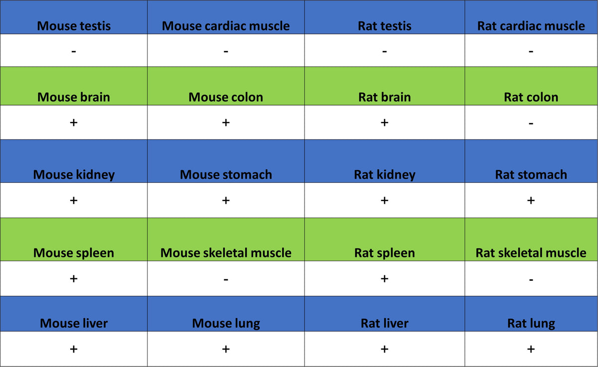
IHC shows positive staining in paraffin-embedded mouse spleen. Anti-Bcl-2 antibody was used at 1/200 dilution, followed by a HRP Polymer for Rabbit IgG (ready to use). Counterstained with hematoxylin. Heat mediated antigen retrieval with Tris/EDTA buffer pH9.0 was performed before commencing with IHC staining protocol.
IHC shows positive staining in paraffin-embedded rat spleen. Anti-Bcl-2 antibody was used at 1/200 dilution, followed by a HRP Polymer for Rabbit IgG (ready to use). Counterstained with hematoxylin. Heat mediated antigen retrieval with Tris/EDTA buffer pH9.0 was performed before commencing with IHC staining protocol.