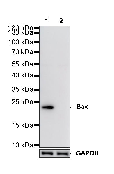
Bax-Knockout HeLa Cell Lysate
WB result:
Primary antibody: Bax Recombinant Rabbit mAb (S0B0230) at 1/1000 dilution
Lane 1: Wild-type HeLa whole cell lysate 20 ug
Lane 2: Bax-Knockout HeLa whole Cell lysate 20 ug
Secondary antibody: Goat Anti-Rabbit lgG (H+L), HRP conjugated at 1/10000 dilution
Predicted band size: 21 kDa
Observed band size: 21 kDa
Bax-Knockout HeLa Cell Lysate
Bax-Knockout HeLa Cell Lysate
Price:
Regular price
$665 USD
Regular price
Sale price
$665 USD
Unit price
per
For shipping services or bulk orders, you may request a quotation.
Secure checkout with

View full details
Product Details
Product Details
Product Specification
| Host | Human |
| Mutations | Mutation in exon3 |
| Application | WB |
| Concentration | 2 mg/ml |
| Physical Appearance | Liquid |
| Storage Buffer | Supplied in SDS Sample Buffer: 50 mM Tris-HCl (pH 6.8 at 25°C), 2% w/v SDS, 10% glycerol, 1% v/v β-Mercaptoethanol, 0.01% w/v bromophenol blue or phenol red. |
| Stability & Storage | -20 °C for 1 months under sterile conditions; |
Background
Bax (Bcl-2-associated X protein) is a pro-apoptotic member of the Bcl-2 protein family and a key regulator of the intrinsic, mitochondria-mediated apoptotic pathway. In response to cellular stress, Bax undergoes conformational activation and translocates from the cytosol to the mitochondrial outer membrane, where it oligomerizes with other pro-apoptotic Bcl-2 family proteins to promote mitochondrial outer membrane permeabilization and the release of cytochrome c. Cytochrome c subsequently contributes to apoptosome formation and activation of caspase-9 and downstream executioner caspases, including caspase-3, ultimately leading to apoptotic cell death. Bax antibodies are widely used in Western blotting, immunohistochemistry, immunofluorescence, and flow cytometry to assess Bax expression, subcellular localization, and apoptosis-related signaling. Caspase-3 knockout may inhibit or delay the downstream execution phase of apoptosis and increase cellular resistance to apoptosis-inducing stimuli, while Bax activation and mitochondrial apoptotic signaling may remain relatively intact, depending on the cell type and apoptotic stimulus.
Picture
Picture
Western Blot