WB result of Aurora A Recombinant Rabbit mAb
Primary antibody: Aurora A Recombinant Rabbit mAb at 1/1000 dilution 4℃ overnight
Lane 1: untreated HT-29 whole cell lysate 20 µg
Lane 2: HT-29 treated with nocodazole (100ng/mL, 16 hours) whole cell lysate 20 µg
Lane 3: untreated HeLa whole cell lysate 20 µg
Lane 4: HeLa treated with nocodazole (100ng/mL, 16 hours) whole cell lysate 20 µg
Secondary antibody: Goat Anti-Rabbit IgG, (H+L), HRP conjugated at 1/10000 dilution
Predicted MW: 46 kDa
Observed MW: 48 kDa
Aurora A Recombinant Rabbit mAb (S-5355)
Aurora A Recombinant Rabbit mAb (S-5355)
Price:
Regular price
$100 USD
Regular price
Sale price
$100 USD
Unit price
per
For shipping services or bulk orders, you may request a quotation.
Secure checkout with

View full details
Product Details
Product Details
Product Specification
| Host | Rabbit |
| Antigen | Aurora A |
| Synonyms | Aurora kinase A; Aurora 2; Aurora/IPL1-related kinase 1 (ARK-1; Aurora-related kinase 1); Breast tumor-amplified kinase; Ipl1- and aurora-related kinase 1; Serine/threonine-protein kinase 15; AIK; AIRK1; ARK1; AURA; AYK1; BTAK; IAK1; STK15 s; STK6; AURKA |
| Location | Cytoplasm, Cytoskeleton |
| Accession | O14965 |
| Clone Number | S-5355 |
| Antibody Type | Recombinant mAb |
| Isotype | IgG |
| Application | WB, IHC-P |
| Reactivity | Hu |
| Purification | Protein A |
| Concentration | 0.5 mg/ml |
| Conjugation | Unconjugated |
| Physical Appearance | Liquid |
| Storage Buffer | PBS, 40% Glycerol, 0.05% BSA, 0.02% sodium azide |
| Stability & Storage | 12 months from date of receipt / reconstitution, -20 °C as supplied |
Dilution
| application | dilution | species |
| WB | 1:1000-1:5000 | Hu |
| IHC-P | 1:500 | Hu |
Background
Aurora A is a serine/threonine kinase that plays a pivotal role in regulating multiple stages of mitosis, including centrosome maturation, spindle assembly, chromosome alignment, and cytokinesis, by phosphorylating key substrates such as TPX2, TACC3, and histone H3 to ensure accurate cell division. Overexpression or amplification of the AURKA gene encoding Aurora A is frequently observed in various human cancers, where it correlates with chromosomal instability, tumor aggressiveness, and poor prognosis, making it an attractive therapeutic target; consequently, numerous small-molecule inhibitors like alisertib have been developed and evaluated in clinical trials, although challenges remain regarding selectivity, resistance mechanisms, and optimal combination strategies to effectively exploit Aurora A dependency in malignancies while minimizing toxicity to normal proliferating tissues.
Picture
Picture
Western Blot
Immunohistochemistry
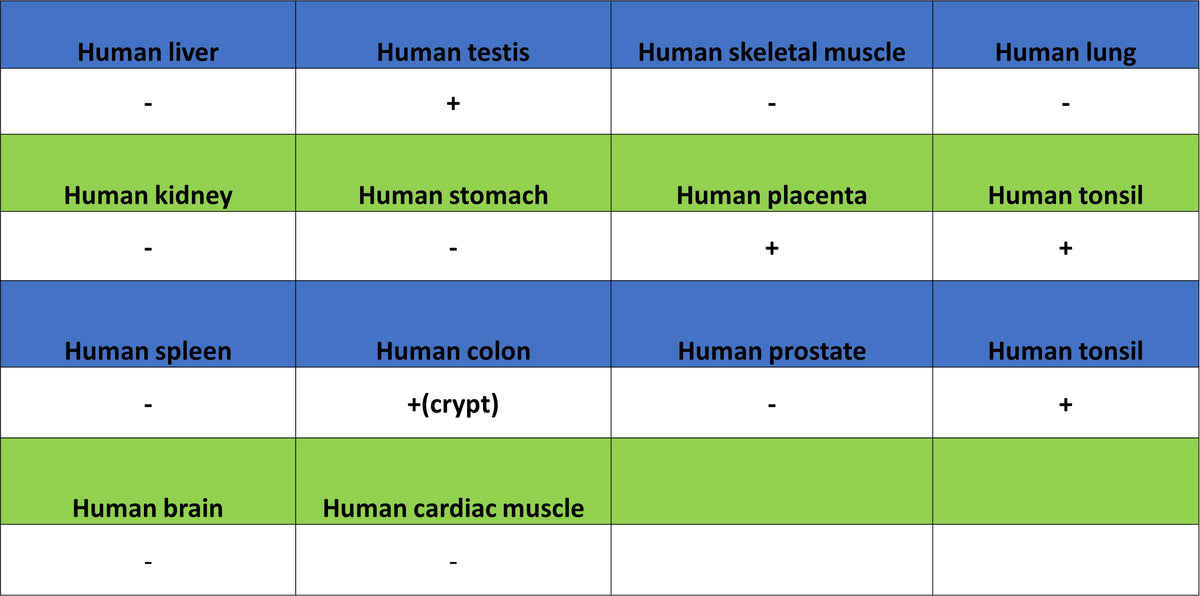
IHC shows positive staining in paraffin-embedded human tonsil. Anti-Aurora A antibody was used at 1/500 dilution, followed by a HRP Polymer for Rabbit IgG (ready to use). Counterstained with hematoxylin. Heat mediated antigen retrieval with Tris/EDTA buffer pH9.0 was performed before commencing with IHC staining protocol.
IHC shows positive staining in paraffin-embedded human gastric cancer. Anti-Aurora A antibody was used at 1/500 dilution, followed by a HRP Polymer for Rabbit IgG (ready to use). Counterstained with hematoxylin. Heat mediated antigen retrieval with Tris/EDTA buffer pH9.0 was performed before commencing with IHC staining protocol.
IHC shows positive staining in paraffin-embedded human lung cancer. Anti-Aurora A antibody was used at 1/500 dilution, followed by a HRP Polymer for Rabbit IgG (ready to use). Counterstained with hematoxylin. Heat mediated antigen retrieval with Tris/EDTA buffer pH9.0 was performed before commencing with IHC staining protocol.