WB result of ATF-6 Recombinant Rabbit mAb
Primary antibody: ATF-6 Recombinant Rabbit mAb at 1/1000 dilution
Lane 1: untreated 293T whole cell lysate 20 µg
Lane 2: 293T treated with 2 µM tunicamycin for 6 hours whole cell lysate 20 µg
Secondary antibody: Goat Anti- rabbit IgG, (H+L), HRP conjugated at 1/10000 dilution
Predicted MW: 75 kDa
Observed MW: 90-100 kDa
ATF-6 Recombinant Rabbit mAb (S-4366)
ATF-6 Recombinant Rabbit mAb (S-4366)
Price:
Regular price
$100 USD
Regular price
Sale price
$100 USD
Unit price
per
For shipping services or bulk orders, you may request a quotation.
Secure checkout with

View full details
Product Details
Product Details
Product Specification
| Host | Rabbit |
| Antigen | ATF-6 |
| Synonyms | Cyclic AMP-dependent transcription factor ATF-6 alpha; ATF6 |
| Location | Nucleus, Endoplasmic reticulum |
| Accession | P18850 |
| Clone Number | S-4366 |
| Antibody Type | Recombinant mAb |
| Isotype | IgG |
| Application | WB, IHC-P |
| Reactivity | Hu |
| Purification | Protein A |
| Concentration | 0.5 mg/ml |
| Conjugation | Unconjugated |
| Physical Appearance | Liquid |
| Storage Buffer | PBS, 40% Glycerol, 0.05% BSA, 0.03% Proclin 300 |
| Stability & Storage | 12 months from date of receipt / reconstitution, -20 °C as supplied |
Dilution
| application | dilution | species |
| WB | 1:1000 | Hu |
| IHC-P | 1:500 | Hu |
Background
ATF-6 protein (activating transcription factor 6) is a key transmembrane protein in the endoplasmic reticulum (ER), belonging to the basic leucine zipper (bZIP) transcription factor family, and plays a central regulatory role in the ER unfolded protein response. Under normal conditions, ATF-6 remains inactive by binding to the molecular chaperone BiP. When the accumulation of misfolded or unfolded proteins in the ER triggers ER stress, BiP dissociates from ATF-6, allowing ATF-6 to translocate to the Golgi apparatus, where it is sequentially cleaved by site-1 protease and site-2 protease, releasing its cytosolic domain—the activated transcription factor form. This activated fragment then enters the nucleus, binds to ER stress response elements, and upregulates the expression of various genes, including those encoding molecular chaperones, foldases, and ER-associated degradation pathway components, thereby enhancing ER protein folding capacity, alleviating stress, and restoring cellular homeostasis. Furthermore, ATF-6 is involved in regulating cell differentiation, lipid metabolism, and antioxidant responses. Dysfunction of ATF-6 is closely associated with various diseases, including diabetes, neurodegenerative diseases (such as Alzheimer's and Parkinson's diseases), and certain cancers. Therefore, ATF-6 is not only a key regulator for maintaining ER functional integrity but also an important potential therapeutic target for these diseases.
Picture
Picture
Western Blot
Immunohistochemistry
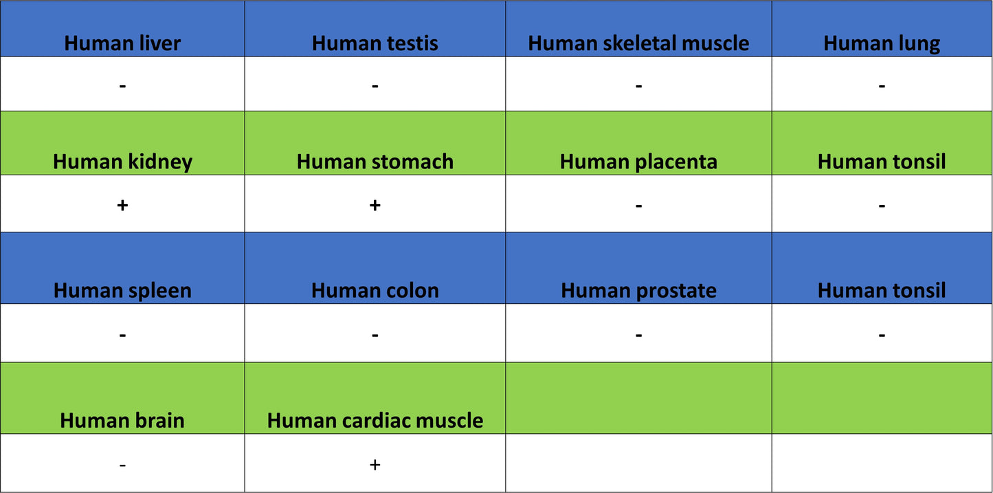
IHC shows positive staining in paraffin-embedded human heart. Anti-ATF-6 antibody was used at 1/500 dilution, followed by a HRP Polymer for Rabbit IgG (ready to use). Counterstained with hematoxylin. Heat mediated antigen retrieval with Tris/EDTA buffer pH9.0 was performed before commencing with IHC staining protocol.
IHC shows positive staining in paraffin-embedded human kidney. Anti-ATF-6 antibody was used at 1/500 dilution, followed by a HRP Polymer for Rabbit IgG (ready to use). Counterstained with hematoxylin. Heat mediated antigen retrieval with Tris/EDTA buffer pH9.0 was performed before commencing with IHC staining protocol.
IHC shows positive staining in paraffin-embedded human stomach. Anti-ATF-6 antibody was used at 1/500 dilution, followed by a HRP Polymer for Rabbit IgG (ready to use). Counterstained with hematoxylin. Heat mediated antigen retrieval with Tris/EDTA buffer pH9.0 was performed before commencing with IHC staining protocol.