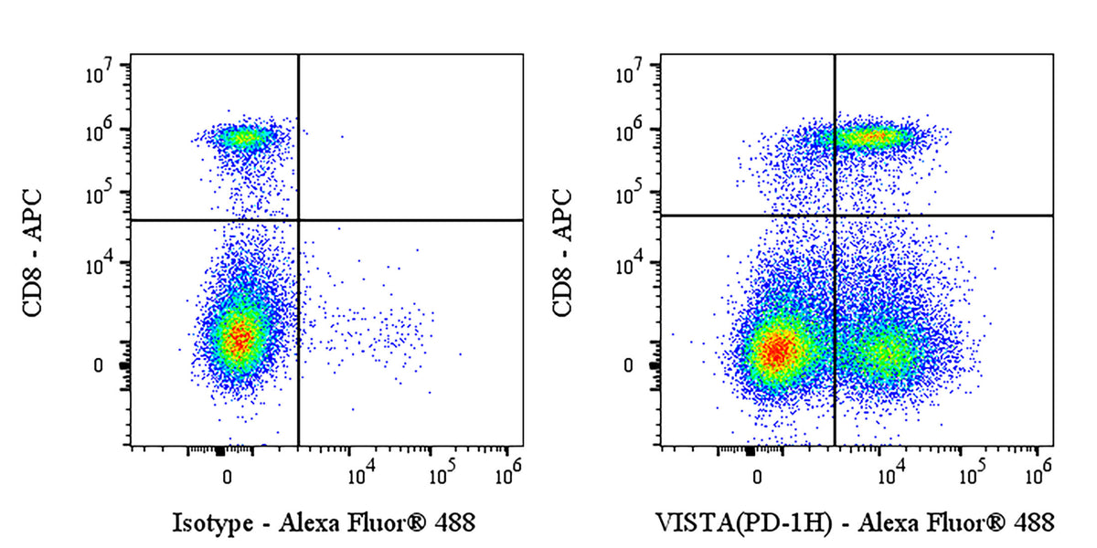
Flow cytometric analysis of C57BL/6 mouse splenocytes labeled with Mouse VISTA antibody at 1/200 dilution (1 μg) / (right panel) compared with an Armenian hamster IgG Isotype Control / (left panel). Goat Anti- Armenian hamster IgG Alexa Fluor® 488 was used as the secondary antibody. Then cells were stained with CD8 – APC antibody separately. Flow cytometry and data analysis were performed using Agilent NovoCyte Quanteon and FlowJo™ software.
Armenian hamster Anti-Mouse VISTA (PD-1H) Antibody (S-R596)
Armenian hamster Anti-Mouse VISTA (PD-1H) Antibody (S-R596)
Price:
Regular price
$135 USD
Regular price
Sale price
$135 USD
Unit price
per
For shipping services or bulk orders, you may request a quotation.
Secure checkout with

View full details
Product Details
Product Details
Product Specification
| Host | Armenian hamster |
| Antigen | VISTA |
| Synonyms | V-type immunoglobulin domain-containing suppressor of T-cell activation; Platelet receptor Gi24; V-set domain-containing immunoregulatory receptorImported; V-set immunoregulatory receptorImported; Dies1; PD-1H; Vsir |
| Location | Cell membrane |
| Accession | Q9D659 |
| Clone Number | S-R596 |
| Antibody Type | Recombinant mAb |
| Isotype | IgG |
| Application | FCM |
| Reactivity | Ms |
| Positive Sample | C57BL/6 mouse splenocytes |
| Purification | Protein G |
| Concentration | 2 mg/ml |
| Conjugation | Unconjugated |
| Physical Appearance | Liquid |
| Storage Buffer | PBS pH7.4 |
| Stability & Storage | 12 months from date of receipt / reconstitution, 2 to 8 °C as supplied |
Dilution
| application | dilution | species |
| FCM | 1:200 | Ms |
Background
VISTA (V-domain Ig suppressor of T-cell activation), also known as B7-H5, PD-1H, C10orf54, Dies1, Gi24 or SISP1, is a type I transmembrane immune-checkpoint protein of the B7-CD28 family that is highly expressed on myeloid and granulocytic cells and at lower levels on CD4+ and CD8+ T cells; it functions both as an inhibitory receptor and as a ligand on antigen-presenting cells to suppress T-cell activation, maintain peripheral tolerance, modulate innate and adaptive anti-tumour immunity, and may additionally regulate embryonic stem-cell differentiation via BMP4 inhibition and stimulate MMP14-mediated MMP2 activation.
Picture
Picture
FC