WB result of ARMC1 Recombinant Rabbit mAb
Primary antibody: ARMC1 Recombinant Rabbit mAb at 1/1000 dilution
Lane 1: Raji whole cell lysate 20 μg
Lane 2: PC-3 whole cell lysate 20 μg
Lane 3: Hep G2 whole cell lysate 20 μg
Lane 4: Jurkat whole cell lysate 20 μg
Lane 5: THP-1 whole cell lysate 20 μg
Lane 6: HL-60 whole cell lysate 20 μg
Lane 7: HT-29 whole cell lysate 20 μg
Lane 8: MDA-MB-231 whole cell lysate 20 μg
Secondary antibody: Goat Anti-Rabbit IgG (H+L), HRP conjugated at 1/10000 dilution
Predicted MW: 31 kDa
Observed MW: 38 kDa
ARMC1 Recombinant Rabbit mAb (S-5206-75)
ARMC1 Recombinant Rabbit mAb (S-5206-75)
Price:
Regular price
$100 USD
Regular price
Sale price
$100 USD
Unit price
per
For shipping services or bulk orders, you may request a quotation.
Secure checkout with

View full details
Product Details
Product Details
Product Specification
| Host | Rabbit |
| Antigen | ARMC1 |
| Immunogen | Synthetic Peptide |
| Location | Cytoplasm, Mitochondrion |
| Accession | Q9NVT9 |
| Clone Number | S-5206-75 |
| Antibody Type | Recombinant mAb |
| Isotype | IgG |
| Application | WB, IHC-P, ICC, IF |
| Reactivity | Hu, Ms, Rt |
| Positive Sample | Raji, PC-3, Hep G2, Jurkat, THP-1, HL-60, HT-29, MDA-MB-231, mouse kidney, mouse liver, rat kidney, rat liver |
| Purification | Protein A |
| Concentration | 0.5 mg/ml |
| Conjugation | Unconjugated |
| Physical Appearance | Liquid |
| Storage Buffer | PBS, 40% Glycerol, 0.05% BSA, 0.02% sodium azide |
| Stability & Storage | 12 months from date of receipt / reconstitution, -20 °C as supplied |
Dilution
| application | dilution | species |
| WB | 1:1000-1:2000 | Hu, Ms, Rt |
| IHC-P | 1:500-1:2000 | Hu, Ms, Rt |
| ICC | 1:500 | Hu |
| IF | 1:100 | Ms, Rt |
Background
ARMC1 (Armadillo repeat-containing protein 1) is a protein of approximately 282 amino acids (molecular weight ~31 kDa) encoded by the human ARMC1 gene located at chromosome 8q13.1, characterized by an Armadillo repeat domain and a conserved C-terminal domain. The protein exhibits dual intracellular localization, being present in both the cytoplasm and mitochondria; it is not integrated into the mitochondrial outer membrane but may associate with it through its C-terminal domain. Functionally, ARMC1 has been identified as a peripheral component of the mitochondrial contact site and cristae organizing system (MICOS) and the mitochondrial intermembrane bridge (MIB) complex, interacting with MICOS components (such as IMMT/MIC60) and outer membrane sorting and assembly machinery (SAM) components (such as SAMM50). Knockout studies indicate that ARMC1 has minimal impact on mitochondrial respiratory function or cristae structure, but it does play a role in regulating mitochondrial length, distribution, and motility. In addition, recent studies suggest that ARMC1 may antagonize TRAK2-mediated retrograde mitochondrial transport by assembling the MIRO–MTFR complex, thereby influencing the intracellular spatial distribution of mitochondria.
Picture
Picture
Western Blot
WB result of Calcineurin A Recombinant Rabbit mAb
Primary antibody: Calcineurin A Recombinant Rabbit mAb at 1/1000 dilution
Lane 1: mouse kidney lysate 20 μg
Lane 2: mouse liver lysate 20 μg
Secondary antibody: Goat Anti-Rabbit IgG (H+L), HRP conjugated at 1/10000 dilution
Predicted MW: 31 kDa
Observed MW: 38 kDa
WB result of Calcineurin A Recombinant Rabbit mAb
Primary antibody: Calcineurin A Recombinant Rabbit mAb at 1/1000 dilution
Lane 1: rat kidney lysate 20 μg
Lane 2: rat liver lysate 20 μg
Secondary antibody: Goat Anti-Rabbit IgG (H+L), HRP conjugated at 1/10000 dilution
Predicted MW: 31 kDa
Observed MW: 38 kDa
Immunohistochemistry
IHC shows positive staining in paraffin-embedded human colon. Anti-ARMC1 antibody was used at 1/2000 dilution, followed by a HRP Polymer for Rabbit IgG (ready to use). Counterstained with hematoxylin. Heat mediated antigen retrieval with Tris/EDTA buffer pH9.0 was performed before commencing with IHC staining protocol.
IHC shows positive staining in paraffin-embedded human kidney. Anti-ARMC1 antibody was used at 1/2000 dilution, followed by a HRP Polymer for Rabbit IgG (ready to use). Counterstained with hematoxylin. Heat mediated antigen retrieval with Tris/EDTA buffer pH9.0 was performed before commencing with IHC staining protocol.
IHC shows positive staining in paraffin-embedded human stomach. Anti-ARMC1 antibody was used at 1/2000 dilution, followed by a HRP Polymer for Rabbit IgG (ready to use). Counterstained with hematoxylin. Heat mediated antigen retrieval with Tris/EDTA buffer pH9.0 was performed before commencing with IHC staining protocol.
IHC shows positive staining in paraffin-embedded human colon cancer. Anti-ARMC1 antibody was used at 1/500 dilution, followed by a HRP Polymer for Rabbit IgG (ready to use). Counterstained with hematoxylin. Heat mediated antigen retrieval with Tris/EDTA buffer pH9.0 was performed before commencing with IHC staining protocol.
IHC shows positive staining in paraffin-embedded human ovarian cancer. Anti-ARMC1 antibody was used at 1/500 dilution, followed by a HRP Polymer for Rabbit IgG (ready to use). Counterstained with hematoxylin. Heat mediated antigen retrieval with Tris/EDTA buffer pH9.0 was performed before commencing with IHC staining protocol.
IHC shows positive staining in paraffin-embedded human gastric cancer. Anti-ARMC1 antibody was used at 1/500 dilution, followed by a HRP Polymer for Rabbit IgG (ready to use). Counterstained with hematoxylin. Heat mediated antigen retrieval with Tris/EDTA buffer pH9.0 was performed before commencing with IHC staining protocol.
IHC shows positive staining in paraffin-embedded mouse brain. Anti-ARMC1 antibody was used at 1/500 dilution, followed by a HRP Polymer for Rabbit IgG (ready to use). Counterstained with hematoxylin. Heat mediated antigen retrieval with Tris/EDTA buffer pH9.0 was performed before commencing with IHC staining protocol.
IHC shows positive staining in paraffin-embedded rat kidney. Anti-ARMC1 antibody was used at 1/500 dilution, followed by a HRP Polymer for Rabbit IgG (ready to use). Counterstained with hematoxylin. Heat mediated antigen retrieval with Tris/EDTA buffer pH9.0 was performed before commencing with IHC staining protocol.
Immunocytochemistry
ICC shows positive staining in Raji cells. Anti-ARMC1 antibody was used at 1/500 dilution (Green) and incubated overnight at 4°C. Goat polyclonal Antibody to Rabbit IgG - H&L (Alexa Fluor® 488) was used as secondary antibody at 1/1000 dilution. The cells were fixed with 4% PFA and permeabilized with 0.1% PBS-Triton X-100. Nuclei were counterstained with DAPI (Blue). Counterstain with tubulin (Red).
Immunofluorescence
IF shows positive staining in paraffin-embedded mouse liver. Anti-ARMC1 antibody was used at 1/100 dilution (Green) and incubated overnight at 4°C. Goat polyclonal Antibody to Rabbit IgG - H&L (Alexa Fluor® 488) was used as secondary antibody at 1/1000 dilution. Counterstained with DAPI (Blue). Heat mediated antigen retrieval with EDTA buffer pH9.0 was performed before commencing with IF staining protocol.
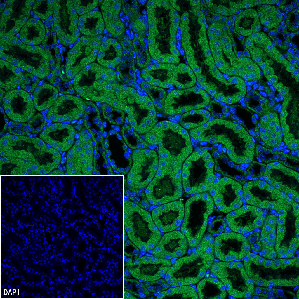
IF shows positive staining in paraffin-embedded mouse kidney. Anti-ARMC1 antibody was used at 1/100 dilution (Green) and incubated overnight at 4°C. Goat polyclonal Antibody to Rabbit IgG - H&L (Alexa Fluor® 488) was used as secondary antibody at 1/1000 dilution. Counterstained with DAPI (Blue). Heat mediated antigen retrieval with EDTA buffer pH9.0 was performed before commencing with IF staining protocol.
IF shows positive staining in paraffin-embedded rat liver. Anti-ARMC1 antibody was used at 1/100 dilution (Green) and incubated overnight at 4°C. Goat polyclonal Antibody to Rabbit IgG - H&L (Alexa Fluor® 488) was used as secondary antibody at 1/1000 dilution. Counterstained with DAPI (Blue). Heat mediated antigen retrieval with EDTA buffer pH9.0 was performed before commencing with IF staining protocol.
IF shows positive staining in paraffin-embedded rat kidney. Anti-ARMC1 antibody was used at 1/100 dilution (Green) and incubated overnight at 4°C. Goat polyclonal Antibody to Rabbit IgG - H&L (Alexa Fluor® 488) was used as secondary antibody at 1/1000 dilution. Counterstained with DAPI (Blue). Heat mediated antigen retrieval with EDTA buffer pH9.0 was performed before commencing with IF staining protocol.