WB result of APP Recombinant Rabbit mAb
Primary antibody: APP Recombinant Rabbit mAb at 1/10000 dilution
Lane 1: 293T whole cell lysate 20 µg
Lane 2: HeLa whole cell lysate 20 µg
Lane 3: U87-MG whole cell lysate 20 µg
Lane 4: SH-SY5Y whole cell lysate 20 µg
Secondary antibody: Goat Anti-Rabbit IgG, (H+L), HRP conjugated at 1/10000 dilution
Predicted MW: 87 kDa
Observed MW: 110-140 kDa
APP Recombinant Rabbit mAb
APP Recombinant Rabbit mAb
Price:
Regular price
$100 USD
Regular price
Sale price
$100 USD
Unit price
per
For shipping services or bulk orders, you may request a quotation.
Secure checkout with

View full details
Product Details
Product Details
Product Specification
| Host | Rabbit |
| Antigen | APP |
| Synonyms | Amyloid-beta precursor protein; APP; ABPP; APPI; Alzheimer disease amyloid A4 protein homolog; Alzheimer disease amyloid protein; Amyloid precursor protein; A4; AD1 |
| Location | Cell membrane |
| Accession | P05067 |
| Antibody Type | Recombinant mAb |
| Isotype | IgG |
| Application | WB, IHC-P |
| Reactivity | Hu, Ms, Rt |
| Positive Sample | 293T, HeLa, U87-MG, SH-SY5Y, 3T3-L1, mouse brain, C6, rat brain |
| Purification | Protein A |
| Concentration | 1 mg/ml |
| Conjugation | Unconjugated |
| Physical Appearance | Liquid |
| Storage Buffer | PBS, 40% Glycerol, 0.05% BSA, 0.02% sodium azide |
| Stability & Storage | 12 months from date of receipt / reconstitution, -20 °C as supplied |
Dilution
| application | dilution | species |
| WB | 1:1000-1:10000 | Hu, Ms, Rt |
| IHC-P | 1:1000 | Hu, Ms, Rt |
Background
Amyloid beta 1-40 (Aβ1-40) is a 40-residue peptide derived from the amyloid precursor protein (APP) through sequential cleavage by β-secretase and γ-secretase. It is one of the major forms of amyloid beta peptides, along with Aβ1-42, and constitutes a significant portion of the amyloid plaques found in the brains of Alzheimer's disease patients. Unlike its more pathogenic counterpart Aβ1-42, Aβ1-40 is considered less prone to aggregation and plaque formation. However, it still plays a role in the disease pathology and has been implicated in neuroinflammation and cerebrovascular dysfunction. Additionally, Aβ1-40 has been found to have non-pathological functions and is more abundant in the brain compared to Aβ1-42.
Picture
Picture
Western Blot
WB result of APP Recombinant Rabbit mAb
Primary antibody: APP Recombinant Rabbit mAb at 1/10000 dilution
Lane 1: 3T3-L1 whole cell lysate 20 µg
Lane 2: mouse brain lysate 20 µg
Secondary antibody: Goat Anti-Rabbit IgG, (H+L), HRP conjugated at 1/10000 dilution
Predicted MW: 87 kDa
Observed MW: 110-130 kDa
WB result of APP Recombinant Rabbit mAb
Primary antibody: APP Recombinant Rabbit mAb at 1/10000 dilution
Lane 1: C6 whole cell lysate 20 µg
Lane 2: rat brain lysate 20 µg
Secondary antibody: Goat Anti-Rabbit IgG, (H+L), HRP conjugated at 1/10000 dilution
Predicted MW: 87 kDa
Observed MW: 110-140 kDa
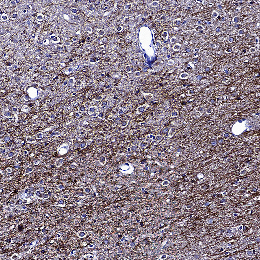
Immunohistochemistry
IHC shows positive staining in paraffin-embedded human brain. Anti-APP antibody was used at 1/1000 dilution, followed by a HRP Polymer for Rabbit IgG (ready to use). Counterstained with hematoxylin. Heat mediated antigen retrieval with Tris/EDTA buffer pH9.0 was performed before commencing with IHC staining protocol.
IHC shows positive staining in paraffin-embedded mouse brain. Anti-APP antibody was used at 1/1000 dilution, followed by a HRP Polymer for Rabbit IgG (ready to use). Counterstained with hematoxylin. Heat mediated antigen retrieval with Tris/EDTA buffer pH9.0 was performed before commencing with IHC staining protocol.
IHC shows positive staining in paraffin-embedded rat brain. Anti-APP antibody was used at 1/1000 dilution, followed by a HRP Polymer for Rabbit IgG (ready to use). Counterstained with hematoxylin. Heat mediated antigen retrieval with Tris/EDTA buffer pH9.0 was performed before commencing with IHC staining protocol.