WB result of APOL2 Recombinant Rabbit mAb
Primary antibody: APOL2 Recombinant Rabbit mAb at 1/1000 dilution
Lane 1: THP-1 whole cell lysate 20 µg
Lane 2: SK-OV-3 whole cell lysate 20 µg
Lane 3: Saos-2 whole cell lysate 20 µg
Low expression control: THP-1 whole cell lysate
Secondary antibody: Goat Anti- rabbit IgG, (H+L), HRP conjugated at 1/10000 dilution
Predicted MW: 37 kDa
Observed MW: 37 kDa
APOL2 Recombinant Rabbit mAb (S-3627-115)
APOL2 Recombinant Rabbit mAb (S-3627-115)
Price:
Regular price
$100 USD
Regular price
Sale price
$100 USD
Unit price
per
For shipping services or bulk orders, you may request a quotation.
Secure checkout with

View full details
Product Details
Product Details
Product Specification
| Host | Rabbit |
| Antigen | APOL2 |
| Synonyms | Apolipoprotein L2 |
| Immunogen | Synthetic Peptide |
| Location | Cytoplasm |
| Accession | Q9BQE5 |
| Clone Number | S-3627-115 |
| Antibody Type | Recombinant mAb |
| Isotype | IgG |
| Application | WB, IHC-P |
| Reactivity | Hu |
| Positive Sample | SK-OV-3, Saos-2, RPMI-8226 |
| Purification | Protein A |
| Concentration | 0.5 mg/ml |
| Conjugation | Unconjugated |
| Physical Appearance | Liquid |
| Storage Buffer | PBS, 40% Glycerol, 0.05% BSA, 0.03% Proclin 300 |
| Stability & Storage | 12 months from date of receipt / reconstitution, -20 °C as supplied |
Dilution
| application | dilution | species |
| WB | 1:1000 | Hu |
| IHC-P | 1:250 | Hu |
Background
Apolipoprotein L2 (APOL2) is a member of the apolipoprotein L family, primarily synthesized in the liver and secreted into the bloodstream as a component of high-density lipoprotein (HDL) particles, where it plays a significant role in lipid metabolism and reverse cholesterol transport. Structurally, APOL2 shares homology with other APOL proteins, containing an N-terminal signal peptide, a central membrane-associating domain, and a C-terminal SRA-interacting domain, although unlike its paralog APOL1, it does not exhibit trypanolytic activity against Trypanosoma brucei rhodesiense. Beyond its metabolic functions, emerging research suggests that APOL2 may be involved in cellular processes such as autophagy and inflammation, potentially interacting with intracellular membranes to influence vesicle trafficking or serving as a biomarker in certain pathological conditions including kidney disease and cancer, though its precise mechanistic contributions to these non-lipid pathways remain an active area of investigation compared to the more extensively characterized roles of other family members.
Picture
Picture
Western Blot
WB result of APOL2 Recombinant Rabbit mAb
Primary antibody: APOL2 Recombinant Rabbit mAb at 1/1000 dilution
Lane 1: RPMI-8226 whole cell lysate 20 µg
Secondary antibody: Goat Anti- rabbit IgG, (H+L), HRP conjugated at 1/10000 dilution
Predicted MW: 37 kDa
Observed MW: 37 kDa
Immunohistochemistry
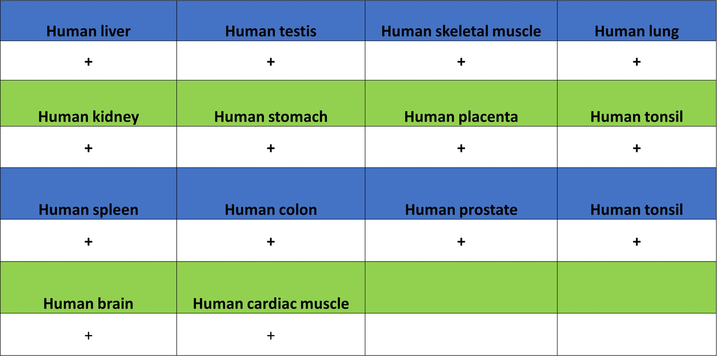
IHC shows positive staining in paraffin-embedded human liver. Anti-APOL2 antibody was used at 1/250 dilution, followed by a HRP Polymer for Rabbit IgG (ready to use). Counterstained with hematoxylin. Heat mediated antigen retrieval with Tris/EDTA buffer pH9.0 was performed before commencing with IHC staining protocol.
IHC shows positive staining in paraffin-embedded human placenta. Anti-APOL2 antibody was used at 1/250 dilution, followed by a HRP Polymer for Rabbit IgG (ready to use). Counterstained with hematoxylin. Heat mediated antigen retrieval with Tris/EDTA buffer pH9.0 was performed before commencing with IHC staining protocol.
IHC shows positive staining in paraffin-embedded human testis. Anti-APOL2 antibody was used at 1/250 dilution, followed by a HRP Polymer for Rabbit IgG (ready to use). Counterstained with hematoxylin. Heat mediated antigen retrieval with Tris/EDTA buffer pH9.0 was performed before commencing with IHC staining protocol.