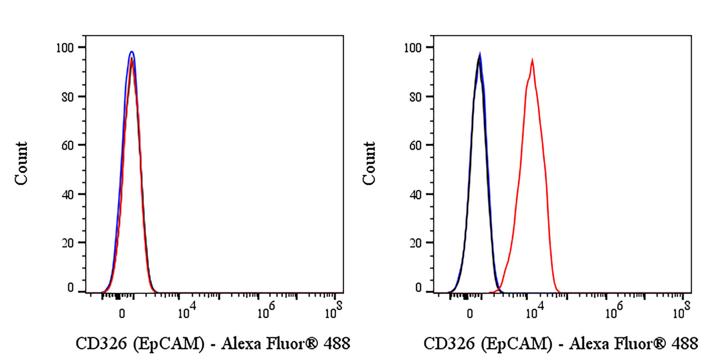
Flow cytometric analysis of HeLa (Human cervix adenocarcinoma epithelial cell, left) / HT29 (Human colorectal adenocarcinoma epithelial cell, right) labeled with CD326 antibody at 1/1000 dilution (0.5 μg) / (red) compared with a Mouse IgG2a,κ (black) Isotype Control and an unlabelled control (cells without incubation with primary antibody and secondary antibody) (blue). Goat Anti - Mouse IgG Alexa Fluor® 488 was used as the secondary antibody. Total viable cells, as determined by Fixable Viability Dye 583 (S0B88803), were used for analysis. Flow cytometry and data analysis were performed using Agilent NovoCyte Quanteon and FlowJo™ software.
Negative control: HeLa
Anti-Human CD326 (EpCAM) Monclonal Antibody (Edrecolomab)
Anti-Human CD326 (EpCAM) Monclonal Antibody (Edrecolomab)
Price:
Regular price
$250 USD
Regular price
Sale price
$250 USD
Unit price
per
For shipping services or bulk orders, you may request a quotation.
Secure checkout with

View full details
Product Details
Product Details
Product Specification
| Host | Mouse |
| Antigen | EpCAM |
| Synonyms | Epithelial cell adhesion molecule; Ep-CAM; Adenocarcinoma-associated antigen; Cell surface glycoprotein Trop-1; Epithelial cell surface antigen; Epithelial glycoprotein (EGP); Epithelial glycoprotein 314 (EGP314; hEGP314); GA733-2; M1S2; M4S1; MIC18; TACSTD1; TROP1; EPCAM |
| Location | Cell membrane |
| Accession | P16422 |
| Clone Number | S-4670 |
| Antibody Type | Mouse mAb |
| Isotype | IgG2a,k |
| Application | Blocking of CD326 signaling Functional assays |
| Reactivity | Hu |
| Positive Sample | HT29 |
| Purification | Protein A |
| Concentration | 5 mg/ml |
| Endotoxin | <1EU/mg |
| Conjugation | Unconjugated |
| Physical Appearance | Liquid |
| Storage Buffer | PBS pH7.4, containing no preservative |
| Stability & Storage | 2 to 8 °C for 2 weeks under sterile conditions; |
Dilution
| application | dilution | species |
| FCM | 1:1000 | Hu |
Background
EpCAM (epithelial cell adhesion molecule, CD326) is a 35–40 kDa type-I transmembrane glycoprotein expressed on most epithelia, where it first assembles into cis-dimers that further cluster into tetramers to support homotypic cell–cell contacts, and then—through sequential proteolytic cleavage—releases its intracellular domain (EpICD) that partners with β-catenin, FHL2 and LEF1 to enter the nucleus and drive Wnt-dependent transcription of genes controlling proliferation, stemness and epithelial-to-mesenchymal transition, thereby functioning not merely as an adhesion molecule but as a dynamic signaling hub that is highly up-regulated in most carcinomas, marks circulating tumor and cancer stem cells, and serves as both a prognostic biomarker and a therapeutic target while mutations that truncate or mis-localize the protein underlie the severe congenital diarrhea disorder tufting enteropathy.
Picture
Picture
FC