Anti-GST Tag Acceptor Beads
Anti-GST Tag Acceptor Beads

Product Details
Product Details
Product Specification
| Stability & Storage | Store at 2~8°C away from light; product shelf life is 12 months. |
Background
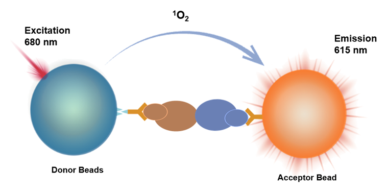
Homogeneous Immuno Chemiluminescence Assay (HICA) is a homogeneous immunoassay method based on energy transfer between donor beads and acceptor beads at close proximity to generate luminescence.
Donor beads recognize protein 1 (Tag1 label), and acceptor beads recognize protein 2 (Tag2 label). When protein 1 binds to protein 2, the distance between the beads becomes less than 200nm. Upon excitation at 680nm, the donor beads generate singlet oxygen, which diffuses to the acceptor beads. The acceptor beads then undergo a redox reaction, emitting light at 615nm. The signal intensity is directly proportional to the strength of the protein interaction.
This product features a simple operation process, requires no washing, and offers fast results with high sensitivity. It is capable of detecting weak interactions.
Components
Specification |
Fill Volume |
250 μg |
50 μL |
5 mg |
1 mL |
25 mg |
1 mL x 5 |
Protocol
[Reagents Required]
Name |
Catalog Number |
| Anti-GST Tag Acceptor Beads | UA086102 |
| Streptavidin Donor Beads | UA086104 |
| Universal Buffer 1 | UA086113 |
[Reference for the Assay Procedure]
Assay procedure |
Assay procedure 1 (37℃ rapid assay) |
Assay procedure 2 (room temperature assay) |
Step one: |
4 μL Tag1-M1 + 4 μL Tag2-M2 + 6 μL Acceptor Beads,Protect from light/green light |
4 μL Tag1-M1 + 4 μL Tag2-M2 + 6 μL Acceptor Beads,Protect from light/green light |
Incubation |
37℃ shaking incubation for 20 minutes,Protect from light/green light | Room temperature incubation for 60 minutes,Protect from light/green light |
Step two: |
Add 6 μL Donor Beads,Protect from light/green light |
Add 6 μL Donor Beads,Protect from light/green light |
Incubate |
Shake and incubate at 37°C for 10 minutes,Protect from light/green light |
Incubate at room temperature for 30 minutes,Protect from light/green light |
Read the results |
Instrument reading |
Instrument reading |
[Performance Verification]
•Sample Preparation:
Using Universal Buffer 1, pre-dilute biotinylated GST (Bio-GST) to 8.1 μg/mL (300 nM) as the stock solution, then perform gradient dilutions according to the following scheme:
Number |
Final Concentration (nM) |
Universal Buffer 1 Volume (μL) |
High-Concentration Addition Volume (μL) |
C12 |
1.0E+01 |
270 |
30 μL of stock solution |
C11 |
3.0E+00 |
210 |
90 μL of C12 |
C10 |
1.0E+00 |
180 |
90 μL of C11 |
C9 |
3.0E-01 |
210 |
90μL C10 |
C8 |
1.0E-01 |
180 |
90μL C9 |
C7 |
3.0E-02 |
210 |
90μL C8 |
C6 |
1.0E-02 |
180 |
90μL C7 |
C5 |
3.0E-03 |
210 |
90μL C6 |
C4 |
1.0E-03 |
180 |
90μL C5 |
C3 |
3.0E-04 |
210 |
90μL C4 |
C2 |
1.0E-04 |
180 |
90μL C3 |
C1 |
0 |
180 |
/ |
•Preparation of test reagents:
Name |
Preparation concentration |
Diluent |
| Anti-GST Tag Acceptor Beads | 25 μg/mL |
Universal Buffer 1 |
| Streptavidin Donor Beads | 25 μg/mL |
Universal Buffer 1 |
•Results of the 37℃ incubation mode:
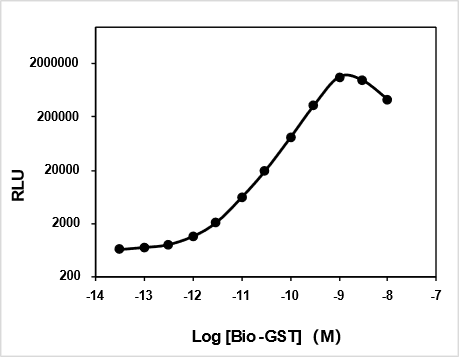
Highest signal: 1,142,982
Lowest signal: 644
EC50 = 0.8679 nM
•Results of the room-temperature incubation mode:
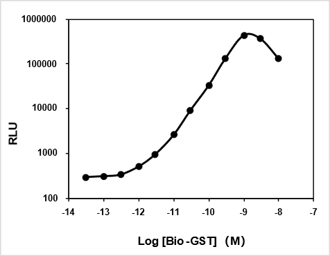
Highest signal: 438,266
Lowest signal: 295
EC50 = 0.8027 nM
Guidelines
1. This experiment is light-sensitive; take care to avoid light during operation. It is recommended to perform the preparation, sample addition, and incubation steps under green light conditions (illuminance below 100 LUX).
2. This product is compatible with multi-mode microplate readers equipped with an Alpha detection module.
3. Before use, vortex thoroughly to mix; a brief centrifugation (2000×g for 5–10 seconds) may also be performed to ensure complete recovery of the reagent.
4. We recommend using our company's dedicated dilution buffer for reagent preparation and sample dilution; if special additives are required, they can be directly added to this dilution buffer.
5. To ensure comparability of experimental data across different batches, strictly control the incubation temperature and time.
6. When adding samples, be careful to avoid introducing air bubbles.