Skip to product information
1 of 12

alpha smooth muscle Actin Recombinant Mouse mAb (SDT-R142)

alpha smooth muscle Actin Recombinant Mouse mAb (SDT-R142)

Catalog Number: S0B0102 Application: WB, IHC-P, ICC, IP, ICFCM Reactivity: Hu, Ms, Rt Conjugation: Brand: Starter
Price:
Regular price $100 USD
Regular price Sale price $100 USD
Size:

For shipping services or bulk orders, you may request a quotation.
Secure checkout with
View full details

Product Details

Product Specification


Host Mouse
Antigen ALPHA SMOOTH MUSCLE ACTIN
Synonyms Alpha-actin-2, ACTA2, SMA
Immunogen N/A
Location Cytoplasm, Cytoskeleton
Accession P62736
Clone Number SDT-R142
Antibody Type Recombinant mAb
Isotype IgG2a
Application WB, IHC-P, ICC, IP, ICFCM
Reactivity Hu, Ms, Rt
Predicted Reactivity Dg
Purification Protein A
Concentration 0.4 mg/ml
Tag N/A
Physical Appearance Liquid
Storage Buffer

PBS, 40% Glycerol, 0.05% BSA, 0.03% Proclin 300

Stability & Storage

12 months from date of receipt / reconstitution, -20 °C as supplied

Dilution


application dilution species
WB 1:1000-1:10000 Ms
IHC-P 1:4000 Hu, Ms, Rt
ICC 1:100 Hu
ICFCM 1:400 Hu
IP 1:40 Ms

Background

α-smooth muscle actin, encoded by ACTA2, is expressed in abundance in vascular smooth muscle, comprising 50–70% of total actin, with the remainder composed of β-cytoplasmic and γ-actins. Whereas α-smooth muscle expression is normally restricted to smooth muscle cells, it can also be expressed in nonmuscle cells, most notably myofibroblasts that use cell traction forces to remodel extracellular matrix.

Picture

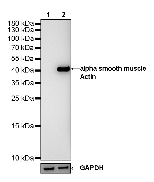
Western Blot

WB result of alpha smooth muscle Actin mouse mAb
Primary antibody: alpha smooth muscle Actin mouse mAb at 1/10000 dilution
Lane 1: mouse skeletal muscle lysate 20 µg
Lane 2: mouse stomach lysate 20 µg
Negative control: mouse skeletal muscle lysate
Secondary antibody: Goat Anti-mouse IgG, (H+L), HRP conjugated at 1/10000 dilution
Predicted MW: 42kDa
Observed MW: 42kDa

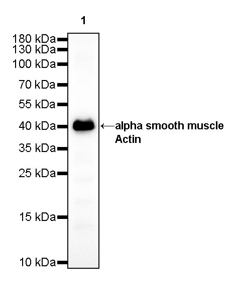
WB result of alpha smooth muscle Actin mouse mAb
Primary antibody: alpha smooth muscle Actin mouse mAb at 1/10000 dilution
Lane 1: NIH/3T3 whole cell lysate 20 µg
Secondary antibody: Goat Anti-mouse IgG, (H+L), HRP conjugated at 1/10000 dilution
Predicted MW: 42kDa
Observed MW: 42kDa

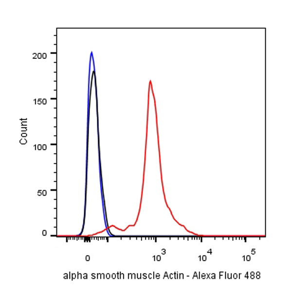
FC

Flow cytometric analysis of HeLa cells labelling alpha smooth muscle Actin antibody at 1/400 (0.1 μg) dilution/ (red) compared with a Mouse monoclonal IgG (Black) isotype control and an unlabelled control (cells without incubation with primary antibody and secondary antibody) (Blue). Goat Anti-Mouse IgG Alexa Fluor® 488 was used as the secondary antibody.

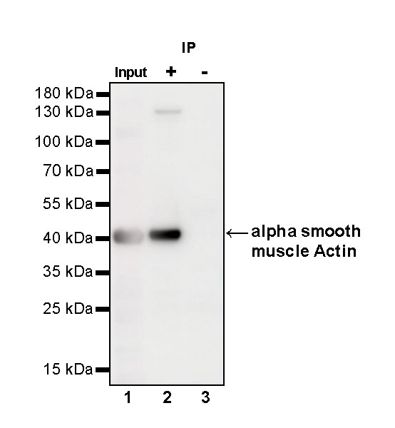
IP

alpha smooth muscle Actin Mouse mAb at 1/40 dilution (1 µg) immunoprecipitating alpha smooth muscle Actin in 0.4 mg NIH/3T3 whole cell lysate.
Western blot was performed on the immunoprecipitate using alpha smooth muscle Actin Mouse mAb at 1/1000 dilution.
Secondary antibody (HRP) for IP was used at 1/400 dilution.
Lane 1: NIH/3T3 whole cell lysate 20 µg (Input)
Lane 2: alpha smooth muscle Actin Mouse mAb IP in NIH/3T3 whole cell lysate
Lane 3: Mouse monoclonal IgG2a IP in NIH/3T3 whole cell lysate
Predicted MW: 42 kDa
Observed MW: 42 kDa


Immunohistochemistry

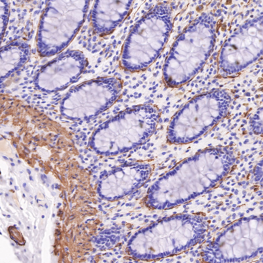
IHC shows positive staining in paraffin-embedded human colon. Anti-alpha smooth muscle Actin antibody was used at 1/4000 dilution, followed by a HRP Polymer for Mouse & Rabbit IgG (ready to use). Counterstained with hematoxylin. Heat mediated antigen retrieval with Tris/EDTA buffer pH9.0 was performed before commencing with IHC staining protocol.

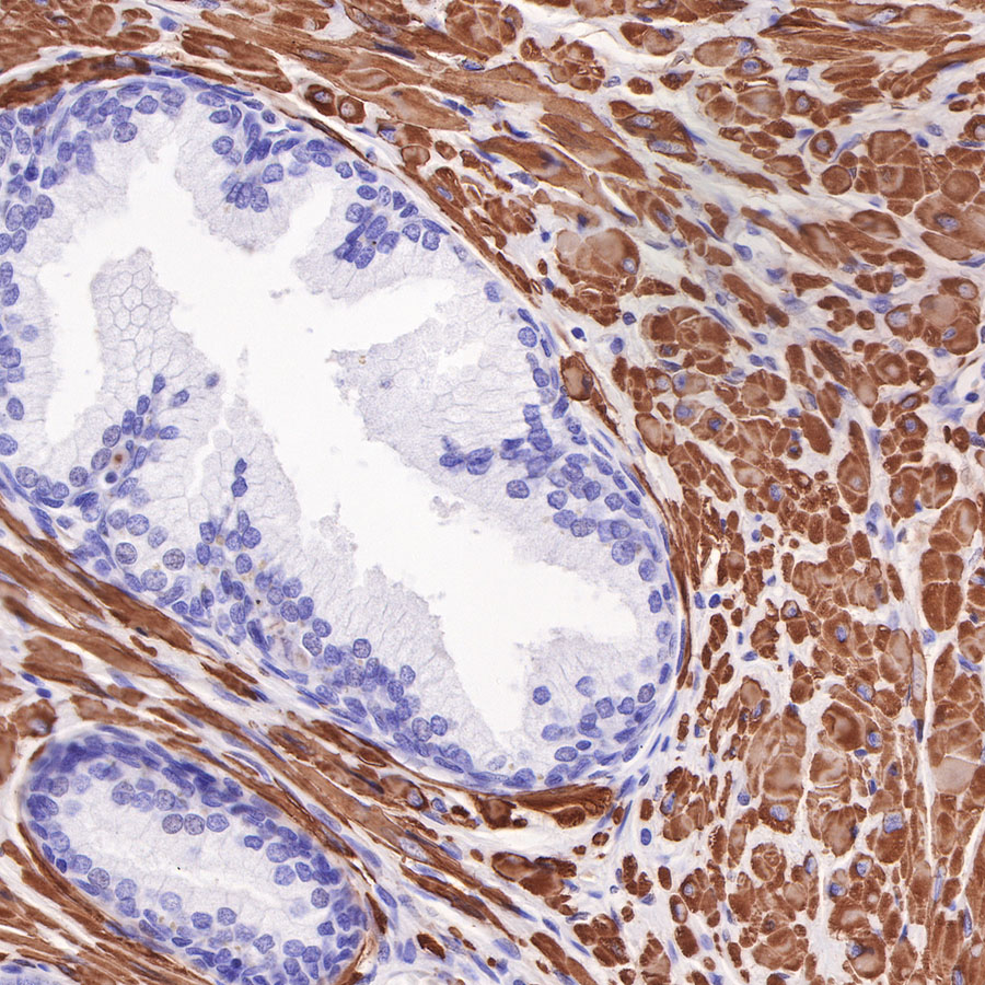
IHC shows positive staining in paraffin-embedded human prostate. Anti-alpha smooth muscle Actin antibody was used at 1/4000 dilution, followed by a HRP Polymer for Mouse & Rabbit IgG (ready to use). Counterstained with hematoxylin. Heat mediated antigen retrieval with Tris/EDTA buffer pH9.0 was performed before commencing with IHC staining protocol.

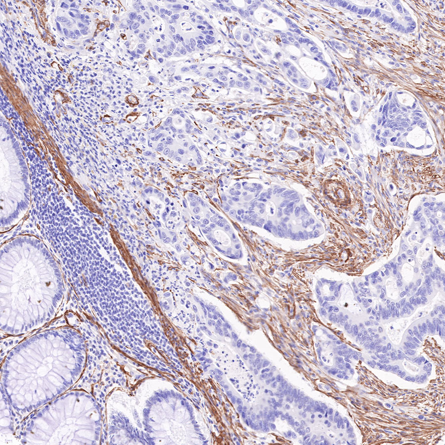
IHC shows positive staining in paraffin-embedded human colon cancer. Anti-alpha smooth muscle Actin antibody was used at 1/4000 dilution, followed by a HRP Polymer for Mouse & Rabbit IgG (ready to use). Counterstained with hematoxylin. Heat mediated antigen retrieval with Tris/EDTA buffer pH9.0 was performed before commencing with IHC staining protocol.

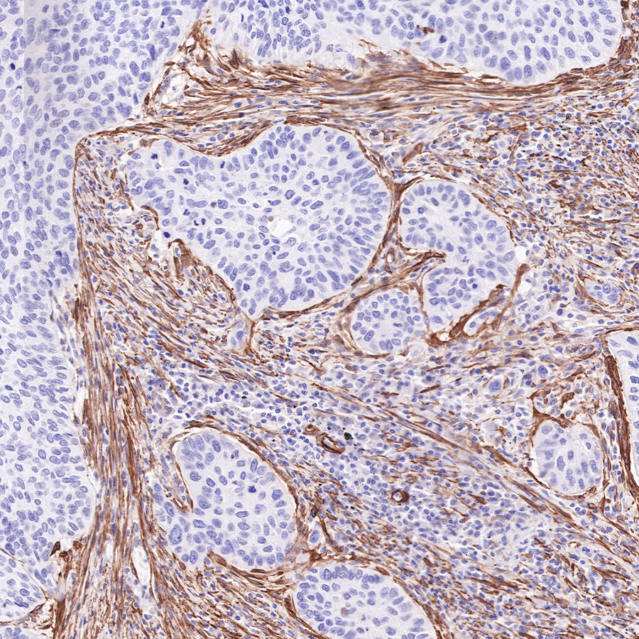
IHC shows positive staining in paraffin-embedded human lung squamous cell carcinoma. Anti-alpha smooth muscle Actin antibody was used at 1/4000 dilution, followed by a HRP Polymer for Mouse & Rabbit IgG (ready to use). Counterstained with hematoxylin. Heat mediated antigen retrieval with Tris/EDTA buffer pH9.0 was performed before commencing with IHC staining protocol.

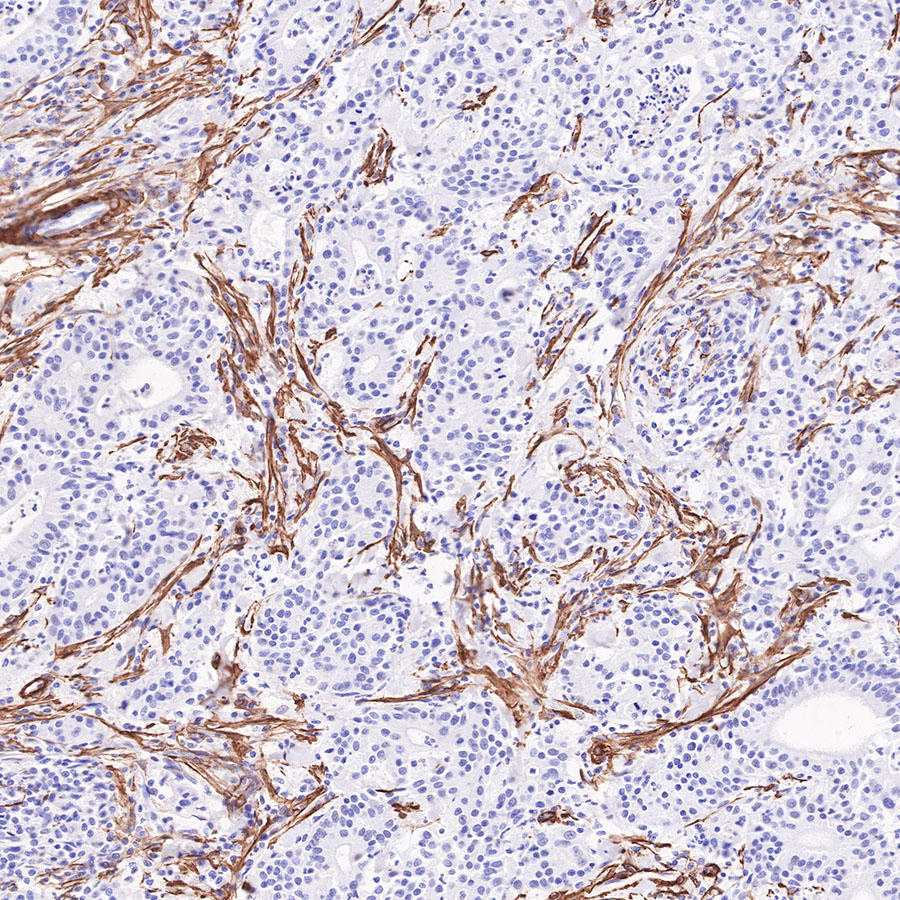
IHC shows positive staining in paraffin-embedded human gastric carcinoma. Anti-alpha smooth muscle Actin antibody was used at 1/4000 dilution, followed by a HRP Polymer for Mouse & Rabbit IgG (ready to use). Counterstained with hematoxylin. Heat mediated antigen retrieval with Tris/EDTA buffer pH9.0 was performed before commencing with IHC staining protocol.

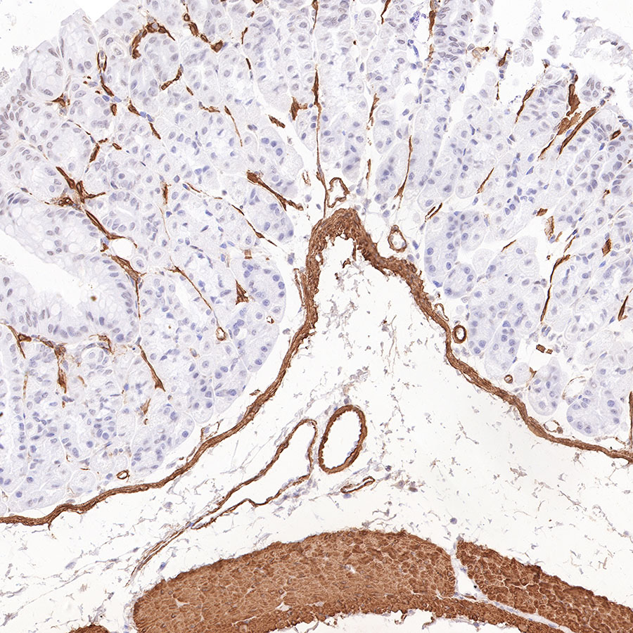
IHC shows positive staining in paraffin-embedded mouse stomach. Anti-alpha smooth muscle Actin antibody was used at 1/4000 dilution, followed by a HRP Polymer for Mouse & Rabbit IgG (ready to use). Counterstained with hematoxylin. Heat mediated antigen retrieval with Tris/EDTA buffer pH9.0 was performed before commencing with IHC staining protocol.

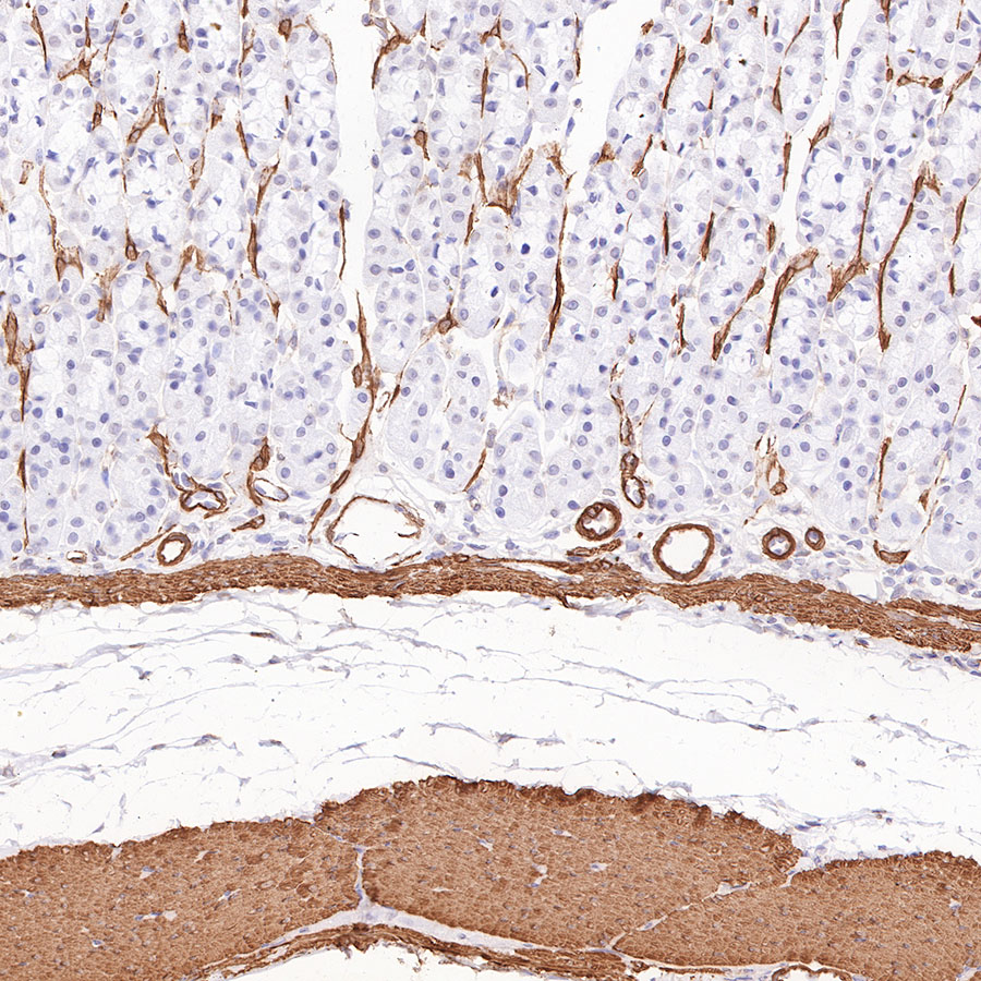
IHC shows positive staining in paraffin-embedded rat stomach. Anti-alpha smooth muscle Actin antibody was used at 1/4000 dilution, followed by a HRP Polymer for Mouse & Rabbit IgG (ready to use). Counterstained with hematoxylin. Heat mediated antigen retrieval with Tris/EDTA buffer pH9.0 was performed before commencing with IHC staining protocol.

Immunocytochemistry

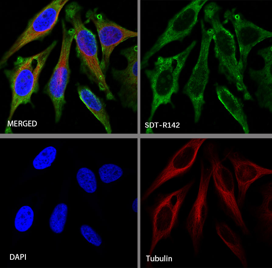
ICC shows positive staining in HeLa cells. Anti-alpha smooth muscle Actin antibody was used at 1/100 dilution and incubated overnight at 4°C. Goat polyclonal Antibody to Mouse IgG - H&L (Alexa Fluor® 488) was used as secondary antibody at 1/1000 dilution. The cells were fixed with 4% PFA and permeabilized with 0.1% PBS-Triton X-100. Nuclei were counterstained with DAPI. Counterstain with tubulin (red).