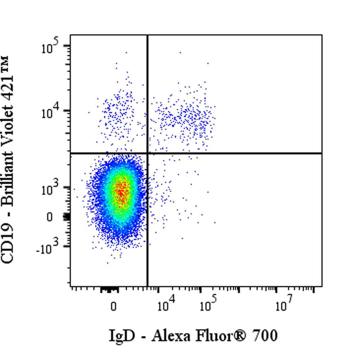
Flow cytometric analysis of Human IgD expression on human peripheral blood leukocytes. Human peripheral blood leukocytes were stained with Brilliant Violet 421™ Mouse Anti-Human CD19 Antibody and Alexa Fluor® 700 Mouse Anti-Human IgD Antibody at 5 μl/test. Flow cytometry and data analysis were performed using Agilent NovoCyte Quanteon and FlowJo™ software.
Alexa Fluor® 700 Mouse Anti-Human IgD Antibody (IA6-2)
Alexa Fluor® 700 Mouse Anti-Human IgD Antibody (IA6-2)
Price:
Regular price
$85 USD
Regular price
Sale price
$85 USD
Unit price
per
For shipping services or bulk orders, you may request a quotation.
Secure checkout with

View full details
Product Details
Product Details
Product Specification
| Host | Mouse |
| Antigen | IgD |
| Synonyms | Immunoglobulin delta heavy chain; Immunoglobulin delta heavy chain WAH |
| Location | Secreted, Cell membrane |
| Accession | P0DOX3 |
| Clone Number | IA6-2 |
| Antibody Type | Mouse mAb |
| Isotype | IgG2a,k |
| Application | FCM |
| Reactivity | Hu |
| Positive Sample | Human peripheral blood leukocytes |
| Purification | Protein A |
| Concentration | 0.2 mg/ml |
| Conjugation | Alexa Fluor® 700 |
| Physical Appearance | Liquid |
| Storage Buffer | PBS, 1% BSA, 0.09% sodium azide |
| Stability & Storage | 12 months from date of receipt / reconstitution, 2 to 8 °C as supplied |
Dilution
| application | dilution | species |
| FCM | 5μl per million cells in 100μl volume | Hu |
Background
Immunoglobulin D (IgD) is an antibody isotype that, in humans, exists primarily as a monomeric, heavily glycosylated membrane-bound receptor on naïve B cells, where it partners with membrane IgM to form the B-cell antigen receptor (BCR) complex that initiates adaptive immune responses upon antigen engagement; it is encoded by the IGH@ locus on chromosome 14, generated through alternative mRNA splicing downstream of IgM, and although secreted IgD is scarce in serum (≈30 µg/mL with a half-life of ~2.8 days), it can be elicited by T-cell–independent stimuli such as interleukin-27 or BAFF and functions through binding to the lectin-like receptor CD44 on basophils and mast cells to trigger antimicrobial and anti-allergic responses, while its hinge region is uniquely elongated and O-glycosylated, imparting flexibility for antigen recognition yet rendering it highly susceptible to proteolysis, and recent evidence indicates that IgD also participates in mucosal immunity via interactions with commensal bacteria and contributes to the development and homeostasis of B-cell subsets, including marginal zone and B-1 cells.
Picture
Picture
FC