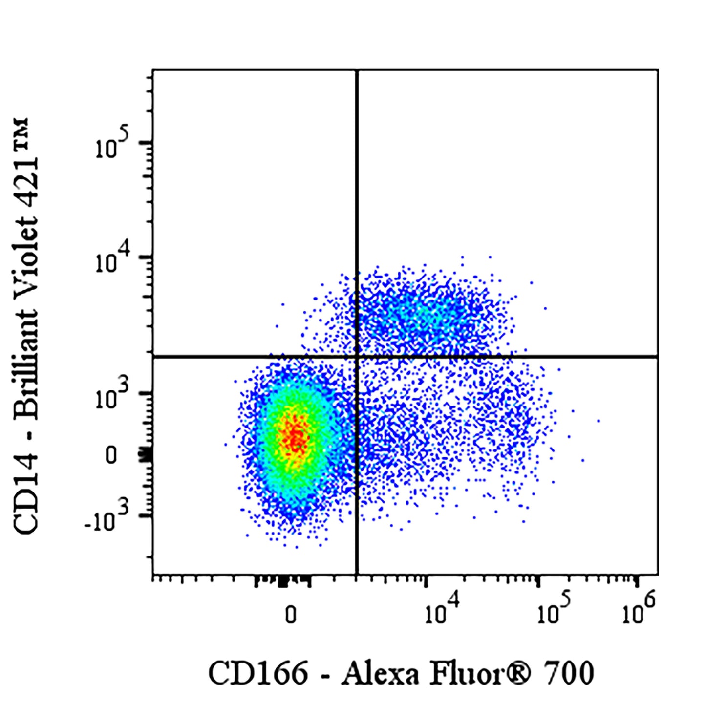
Flow cytometric analysis of human CD166 expression on human PBMCs (human peripheral blood mononuclear cells). Human PBMCs were stained with Brilliant Violet 421™ Mouse Anti-Human CD14 Antibody and SDT Alexa Fluor® 700 Mouse Anti-Human CD166 Antibody at 5 μl/test. Flow cytometry and data analysis were performed using Agilent NovoCyte Quanteon and FlowJo™ software.
Alexa Fluor® 700 Mouse Anti-Human CD166 Antibody (S-2869)
Alexa Fluor® 700 Mouse Anti-Human CD166 Antibody (S-2869)
Price:
Regular price
$120 USD
Regular price
Sale price
$120 USD
Unit price
per
For shipping services or bulk orders, you may request a quotation.
Secure checkout with

View full details
Product Details
Product Details
Product Specification
| Host | Mouse |
| Antigen | CD166 |
| Synonyms | CD166 antigen; Activated leukocyte cell adhesion molecule; CD166; MEMD; ALCAM |
| Location | Cell membrane |
| Accession | Q13740 |
| Clone Number | S-2869 |
| Antibody Type | Mouse mAb |
| Isotype | IgG1,k |
| Application | FCM |
| Reactivity | Hu |
| Positive Sample | Human PBMC |
| Purification | Protein G |
| Concentration | 0.2 mg/ml |
| Conjugation | Alexa Fluor® 700 |
| Physical Appearance | Liquid |
| Storage Buffer | PBS, 1% BSA, 0.09% sodium azide |
| Stability & Storage | 12 months from date of receipt / reconstitution, 2 to 8 °C as supplied |
Dilution
| application | dilution | species |
| FCM | 5μl per million cells in 100μl volume | Hu |
Background
CD166, also known as Activated Leukocyte Cell Adhesion Molecule (ALCAM), is a type I transmembrane glycoprotein belonging to the immunoglobulin superfamily. It is encoded by the ALCAM gene and is involved in both homotypic and heterotypic cell adhesion. CD166 is expressed on various cell types, including activated T cells, epithelial cells, fibroblasts, and neurons. It plays a crucial role in mediating adhesion interactions between thymic epithelial cells and CD6+ cells during T cell development. Additionally, CD166 is overexpressed in several types of tumors and serves as a potential cancer stem cell marker, making it a target for cancer research and therapy.
Picture
Picture
FC