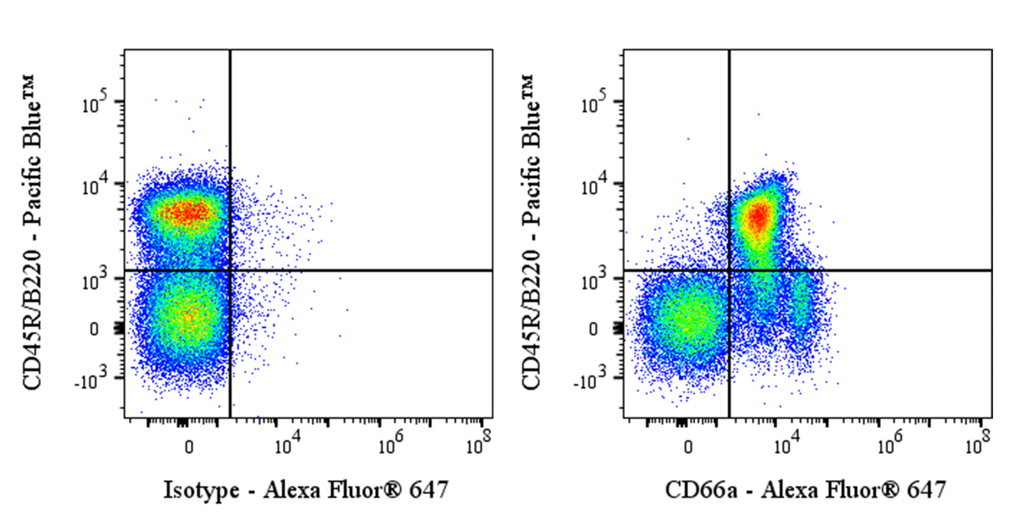
Flow cytometric analysis of mouse CD66a expression on C57BL/6 mouse splenocytes. C57BL/6 mouse splenocytes were stained with Pacific Blue™ Rat Anti-Mouse CD45R/B220 Antibody and either Alexa Fluor® 647 Mouse IgG1, k Isotype Control (left panel) or SDT Alexa Fluor® 647 Mouse Anti-Mouse CD66a Antibody (right panel) at 2 μl/test. Total viable cells, as determined by Fixable Viability Dye 515 (S0D0013), were used for analysis. Flow cytometry and data analysis were performed using Agilent NovoCyte Quanteon and FlowJo™ software.
Alexa Fluor® 647 Mouse Anti-Mouse CD66a Antibody (S-3301)
Alexa Fluor® 647 Mouse Anti-Mouse CD66a Antibody (S-3301)
Price:
Regular price
$170 USD
Regular price
Sale price
$170 USD
Unit price
per
For shipping services or bulk orders, you may request a quotation.
Secure checkout with

View full details
Product Details
Product Details
Product Specification
| Host | Mouse |
| Antigen | CD66a |
| Synonyms | Cell adhesion molecule CEACAM1; Biliary glycoprotein 1 (BGP-1); Biliary glycoprotein D; Carcinoembryonic antigen-related cell adhesion molecule 1 (CEA cell adhesion molecule 1Imported); MHVR1; Murine hepatitis virus receptor (MHV-R); Bgp; Bgp1; Ceacam1 |
| Location | Cell membrane |
| Accession | P31809 |
| Clone Number | S-3301 |
| Antibody Type | Mouse mAb |
| Isotype | IgG1,k |
| Application | FCM |
| Reactivity | Ms |
| Positive Sample | C57BL/6 mouse splenocytes |
| Purification | Protein G |
| Concentration | 0.5 mg/ml |
| Conjugation | Alexa Fluor® 647 |
| Physical Appearance | Liquid |
| Storage Buffer | PBS, 1% BSA, 0.3% Proclin 300 |
| Stability & Storage | 12 months from date of receipt / reconstitution, 2 to 8 °C as supplied |
Dilution
| application | dilution | species |
| FCM | 1μg per million cells in 100μl volume | Ms |
Background
CD66a, also known as CEACAM1, is a transmembrane glycoprotein belonging to the immunoglobulin superfamily that mediates homophilic intercellular adhesion and regulates immune cell activation, apoptosis, and differentiation through phosphatidylinositol 3-kinase- and Akt-dependent survival signaling pathways.
Picture
Picture
FC