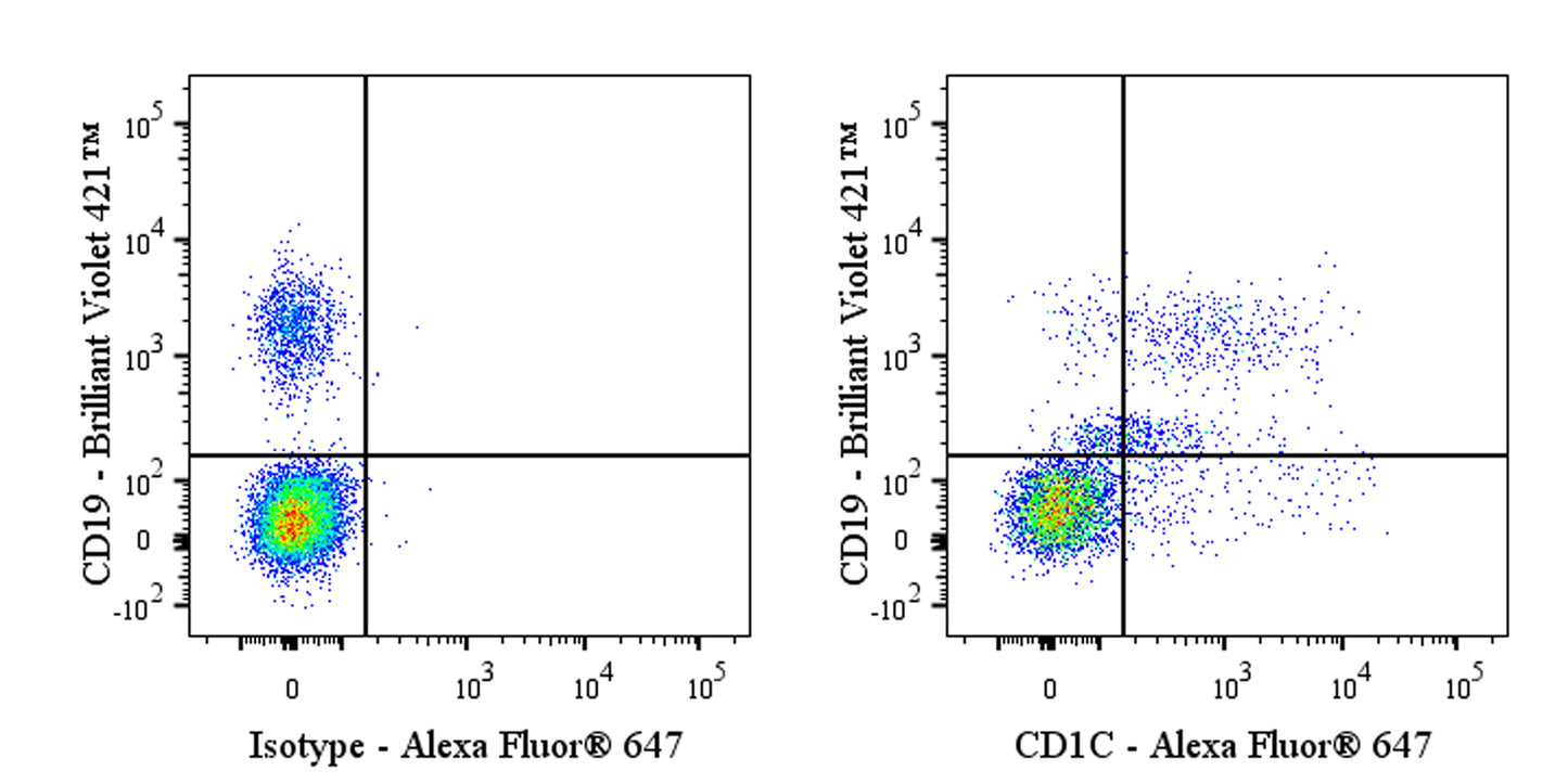
Flow cytometric analysis of CD1C expression on Human PBMC (human peripheral blood mononuclear cells). Human PBMC were stained with Brilliant Violet 421™ Mouse Anti-Human CD19 Antibody and either Alexa Fluor® 647 Mouse IgG1, κ Isotype Control (left panel) or SDT Alexa Fluor® 647 Mouse Anti- Human CD1C Antibody (right panel) at 5 μl/test. Flow cytometry and data analysis were performed using BD FACSymphony™ A1 and FlowJo™ software.
Alexa Fluor® 647 Mouse Anti-Human CD1c Antibody (L616)
Alexa Fluor® 647 Mouse Anti-Human CD1c Antibody (L616)
Price:
Regular price
$100 USD
Regular price
Sale price
$100 USD
Unit price
per
For shipping services or bulk orders, you may request a quotation.
Secure checkout with

View full details
Product Details
Product Details
Product Specification
| Host | Mouse |
| Antigen | CD1c |
| Synonyms | T-cell surface glycoprotein CD1c; CD1C |
| Location | Lysosome, Cell membrane |
| Accession | P29017 |
| Clone Number | L616 |
| Antibody Type | Mouse mAb |
| Isotype | IgG1,k |
| Isotype Control | S0B5964 |
| Application | FCM |
| Reactivity | Hu |
| Positive Sample | Human PBMC |
| Purification | Protein G |
| Concentration | 0.2 mg/ml |
| Conjugation | Alexa Fluor® 647 |
| Physical Appearance | Liquid |
| Storage Buffer | PBS, 1% BSA, 0.3% Proclin 300 |
| Stability & Storage | 12 months from date of receipt / reconstitution, 2 to 8 °C as supplied |
Dilution
| application | dilution | species |
| FCM | 5μl per million cells in 100μl volume | Hu |
Background
CD1c is a lipid-presenting, MHC class I-like glycoprotein expressed on dendritic cells and B cells that binds both self and foreign lipids/glycolipids within its deep hydrophobic groove and shuttles them through endosomal compartments to display them to αβ and γδ T cells, thereby bridging innate and adaptive immunity; uniquely, its open F′ pocket can accommodate both lipid tails and peptide moieties of lipopeptides, enabling recognition of mycobacterial antigens such as mannosyl-β1-phosphomycoketide and driving potent IFN-γ responses essential for controlling intracellular pathogens like Mycobacterium tuberculosis.
Picture
Picture
FC