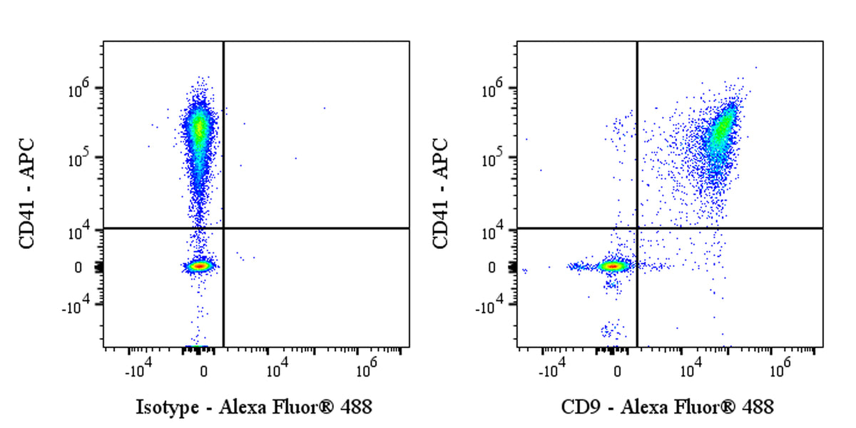
Flow cytometric analysis of CD9 expression on fresh human peripheral blood cells. Fresh human peripheral blood cells were stained with APC Mouse Anti-Human CD41 Antibody and either Alexa Fluor® 488 Rabbit IgG Isotype Control (left panel) or SDT Alexa Fluor® 488 Rabbit Anti-Human CD9 Antibody (right panel) at 5 μl/test. Flow cytometry and data analysis were performed using Agilent NovoCyte Quanteon and FlowJo™ software.
Alexa Fluor® 488 Rabbit Anti-Human CD9 Antibody (S-143-53)
Alexa Fluor® 488 Rabbit Anti-Human CD9 Antibody (S-143-53)
Price:
Regular price
$175 USD
Regular price
Sale price
$175 USD
Unit price
per
For shipping services or bulk orders, you may request a quotation.
Secure checkout with

View full details
Product Details
Product Details
Product Specification
| Host | Rabbit |
| Antigen | CD9 |
| Synonyms | CD9 antigen; 5H9 antigen; Cell growth-inhibiting gene 2 protein; Leukocyte antigen MIC3; Motility-related protein (MRP-1); Tetraspanin-29 (Tspan-29); p24; MIC3; TSPAN29 |
| Immunogen | Recombinant Protein |
| Location | Cell membrane |
| Accession | P21926 |
| Clone Number | S-143-53 |
| Antibody Type | Recombinant mAb |
| Isotype | IgG |
| Isotype Control | S0B0161 |
| Application | FCM |
| Reactivity | Hu |
| Positive Sample | Fresh human peripheral blood |
| Purification | Protein A |
| Concentration | 0.2 mg/ml |
| Conjugation | Alexa Fluor® 488 |
| Physical Appearance | Liquid |
| Storage Buffer | PBS, 1% BSA, 0.09% sodium azide |
| Stability & Storage | 12 months from date of receipt / reconstitution, 2 to 8 °C as supplied |
Dilution
| application | dilution | species |
| FCM | 5μl per million cells in 100μl volume | Hu |
Background
CD9, a 24-kDa cell-surface glycoprotein belonging to the tetraspanin family, is characterized by four transmembrane domains and two extracellular loops; it is widely expressed (e.g., on platelets, B/T cells, macrophages, and oocytes) and organizes tetraspanin-enriched microdomains that integrate membrane and cytoplasmic proteins to regulate adhesion, migration, proliferation, survival, and immune synapse formation. Notably, CD9 is essential for mammalian gamete fusion, as CD9-null eggs cannot fuse with wild-type sperm, and anti-CD9 antibodies block sperm–egg binding and fusion in vitro. Additionally, CD9 is implicated in platelet activation, signal transduction, and is frequently detected in lymphoblastic leukemia/lymphoma and some acute myeloid leukemia cells.
Picture
Picture
FC