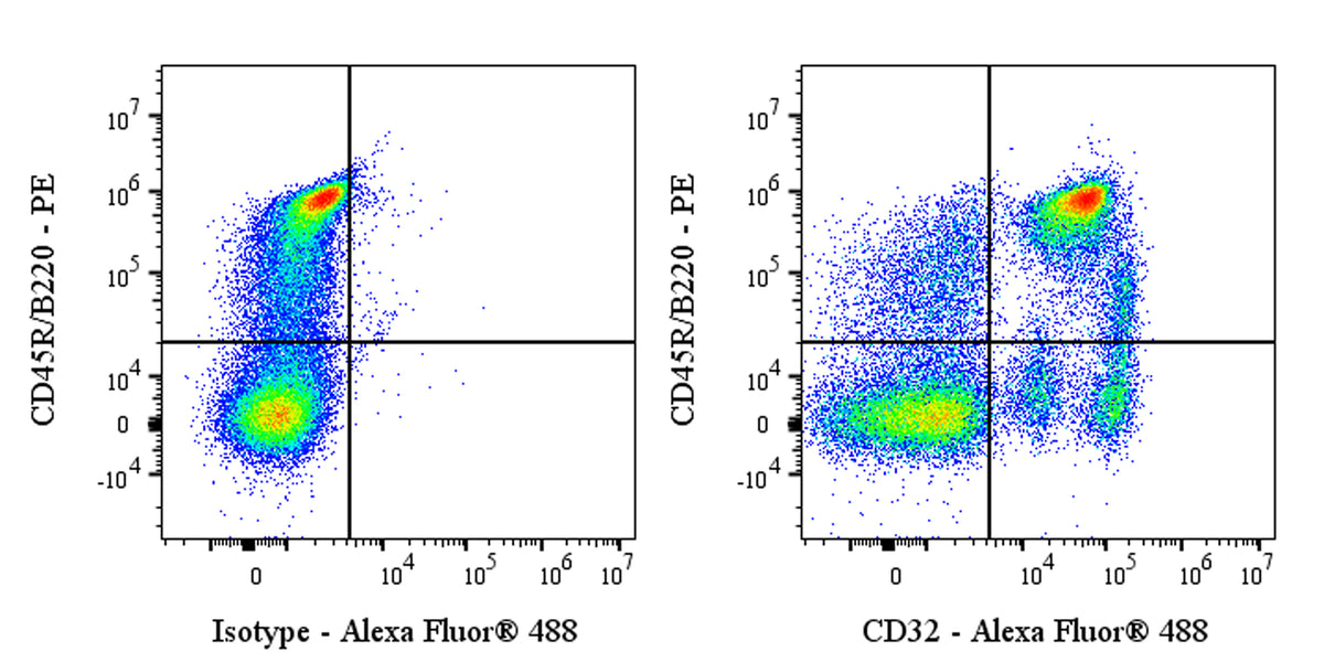
Flow cytometric analysis of Rat CD32 expression on Wister Rat splenocytes. Wister Rat splenocytes were stained with PE Mouse Anti-Rat CD45R/B220 Antibody and either Alexa Fluor® 488 Mouse IgG1, k Isotype Control (left panel) or SDT Alexa Fluor® 488 Mouse Anti-Rat CD32 Antibody (right panel) at 2 μl/test. Total viable cells, as determined by Fixable Viability Dye 452 (S0B88801), were used for analysis. Flow cytometry and data analysis were performed using Agilent NovoCyte Quanteon and FlowJo™ software.
Alexa Fluor® 488 Mouse Anti-Rat CD32 Antibody (S-4246)
Alexa Fluor® 488 Mouse Anti-Rat CD32 Antibody (S-4246)
Price:
Regular price
$130 USD
Regular price
Sale price
$130 USD
Unit price
per
For shipping services or bulk orders, you may request a quotation.
Secure checkout with

View full details
Product Details
Product Details
Product Specification
| Host | Mouse |
| Antigen | CD32 |
| Location | Cell membrane |
| Accession | Q63203 |
| Clone Number | S-4246 |
| Antibody Type | Mouse mAb |
| Isotype Control | S0B1519 |
| Application | FCM |
| Reactivity | Rt |
| Positive Sample | Wister Rat splenocytes |
| Purification | Protein G |
| Concentration | 0.5 mg/ml |
| Conjugation | Alexa Fluor® 488 |
| Physical Appearance | Liquid |
| Storage Buffer | PBS, 1% BSA, 0.09% sodium azide |
| Stability & Storage | 12 months from date of receipt / reconstitution, 2 to 8 °C as supplied |
Dilution
| application | dilution | species |
| FCM | 1μg per million cells in 100μl volume | Rt |
Background
CD32 (FcγRII) is a type I transmembrane glycoprotein of approximately 40 kDa that belongs to the immunoglobulin superfamily and serves as a low- to medium-affinity Fc receptor for IgG, playing a central role in immune regulation. CD32 is encoded by three genes—FCGR2A, FCGR2B, and FCGR2C—and alternative splicing generates at least six isoforms (IIa1, IIa2, IIb1, IIb2, IIb3, and IIc), which differ fundamentally in their intracellular signaling domains. Its extracellular region contains two immunoglobulin-like domains, with the IgG-binding site located mainly in the Asn154-Ser161 segment of the second domain, while other regions of the first and second domains also participate in binding. Functionally, the cytoplasmic tails of CD32A and CD32C contain immunoreceptor tyrosine-based activation motifs (ITAMs); upon ligand binding, they recruit kinases such as Syk and Lyn to trigger activating signals including phagocytosis, antibody-dependent cellular cytotoxicity (ADCC), and cytokine release. CD32B, in contrast, contains an immunoreceptor tyrosine-based inhibitory motif (ITIM) and recruits phosphatases to negatively regulate B cell activation and maintain immune tolerance. CD32 is widely expressed on B cells, monocytes/macrophages, neutrophils, platelets, and placental trophoblasts, and its dysfunction is closely associated with autoimmune diseases such as systemic lupus erythematosus and rheumatoid arthritis.
Picture
Picture
FC