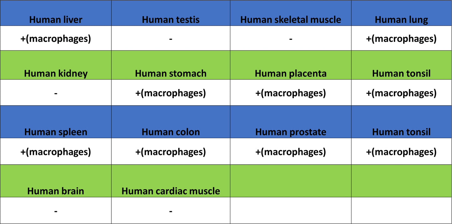
IHC shows positive staining in paraffin-embedded human colon. Anti-ADGRE1 (F4/80) antibody was used at 1/500 dilution, followed by a HRP Polymer for Rabbit IgG (ready to use). Counterstained with hematoxylin. Heat mediated antigen retrieval with Tris/EDTA buffer pH9.0 was performed before commencing with IHC staining protocol.
ADGRE1 (F4/80) Recombinant Rabbit mAb (S-2683-205)
ADGRE1 (F4/80) Recombinant Rabbit mAb (S-2683-205)
Price:
Regular price
$100 USD
Regular price
Sale price
$100 USD
Unit price
per
For shipping services or bulk orders, you may request a quotation.
Secure checkout with

View full details
Product Details
Product Details
Product Specification
| Host | Rabbit |
| Antigen | ADGRE1 (F4/80) |
| Synonyms | Adhesion G protein-coupled receptor E1; EGF-like module receptor 1; EGF-like module-containing mucin-like hormone receptor-like 1; EMR1 hormone receptor; EMR1; TM7LN3 |
| Immunogen | Recombinant Protein |
| Location | Cell membrane |
| Accession | Q14246 |
| Clone Number | S-2683-205 |
| Antibody Type | Recombinant mAb |
| Isotype | IgG |
| Application | IHC-P |
| Reactivity | Hu |
| Purification | Protein A |
| Concentration | 0.5 mg/ml |
| Conjugation | Unconjugated |
| Physical Appearance | Liquid |
| Storage Buffer | PBS, 40% Glycerol, 0.05% BSA, 0.03% Proclin 300 |
| Stability & Storage | 12 months from date of receipt / reconstitution, -20 °C as supplied |
Dilution
| application | dilution | species |
| IHC-P | 1:500 | Hu |
Background
ADGRE1 (Adhesion G protein-coupled receptor E1, also known as EMR1 or F4/80) is a structurally unique macrophage marker membrane protein that plays an important role in immune surveillance and inflammatory regulation. This protein is characterized by a long extracellular N-terminal region containing six epidermal growth factor (EGF)-like modules, connected via a serine/threonine-rich domain to the classic C-terminal seven-transmembrane G protein-coupled receptor domain—a conformation that endows it with dual potential for both cell adhesion and signal transduction. As the most classic macrophage surface marker F4/80 in mice, as well as a functionally conserved receptor in humans, ADGRE1 is primarily localized to the cell membrane and participates in regulating macrophage biological functions, mediating peripheral immune tolerance, and maintaining tissue homeostasis through activation of the adenylate cyclase signaling pathway. Although its specific endogenous ligands remain incompletely defined, studies have shown that this receptor is associated with various disease states, including obesity, fibrosis, and inflammatory bowel disease, highlighting its potential research value in immune-related pathological processes.
Picture
Picture
Immunohistochemistry
IHC shows positive staining in paraffin-embedded human liver. Anti-ADGRE1 (F4/80) antibody was used at 1/500 dilution, followed by a HRP Polymer for Rabbit IgG (ready to use). Counterstained with hematoxylin. Heat mediated antigen retrieval with Tris/EDTA buffer pH9.0 was performed before commencing with IHC staining protocol.
IHC shows positive staining in paraffin-embedded human spleen. Anti-ADGRE1 (F4/80) antibody was used at 1/500 dilution, followed by a HRP Polymer for Rabbit IgG (ready to use). Counterstained with hematoxylin. Heat mediated antigen retrieval with Tris/EDTA buffer pH9.0 was performed before commencing with IHC staining protocol.
IHC shows positive staining in paraffin-embedded human stomach. Anti-ADGRE1 (F4/80) antibody was used at 1/500 dilution, followed by a HRP Polymer for Rabbit IgG (ready to use). Counterstained with hematoxylin. Heat mediated antigen retrieval with Tris/EDTA buffer pH9.0 was performed before commencing with IHC staining protocol.
IHC shows positive staining in paraffin-embedded human lung cancer. Anti-ADGRE1 (F4/80) antibody was used at 1/500 dilution, followed by a HRP Polymer for Rabbit IgG (ready to use). Counterstained with hematoxylin. Heat mediated antigen retrieval with Tris/EDTA buffer pH9.0 was performed before commencing with IHC staining protocol.