WB result of Aass Recombinant Rabbit mAb
Primary antibody: Aass Recombinant Rabbit mAb at 1/1000 dilution
Lane 1: 293T whole cell lysate 20 µg
Lane 2: HepG2 whole cell lysate 20 µg
Lane 3: HUVEC whole cell lysate 20 µg
Lane 4: HeLa whole cell lysate 20 µg
Secondary antibody: Goat Anti- rabbit IgG, (H+L), HRP conjugated at 1/10000 dilution
Predicted MW: 102 kDa
Observed MW: 110 kDa
Aass Recombinant Rabbit mAb (S-4170-22)
Aass Recombinant Rabbit mAb (S-4170-22)
Price:
Regular price
$100 USD
Regular price
Sale price
$100 USD
Unit price
per
For shipping services or bulk orders, you may request a quotation.
Secure checkout with

View full details
Product Details
Product Details
Product Specification
| Host | Rabbit |
| Antigen | Aass |
| Synonyms | Alpha-aminoadipic semialdehyde synthase, mitochondrial; AASS |
| Immunogen | Synthetic Peptide |
| Location | Mitochondrion |
| Accession | Q9UDR5 |
| Clone Number | S-4170-22 |
| Antibody Type | Recombinant mAb |
| Isotype | IgG |
| Application | WB, IHC-P |
| Reactivity | Hu, Ms, Rt |
| Positive Sample | 293T, HepG2, HUVEC, HeLa |
| Purification | Protein A |
| Concentration | 0.5 mg/ml |
| Conjugation | Unconjugated |
| Physical Appearance | Liquid |
| Storage Buffer | PBS, 40% Glycerol, 0.05% BSA, 0.02% sodium azide |
| Stability & Storage | 12 months from date of receipt / reconstitution, -20 °C as supplied |
Dilution
| application | dilution | species |
| WB | 1:1000 | Hu |
| IHC-P | 1:500-1:2000 | Hu, Ms, Rt |
Background
Aass (alpha-aminoadipic semialdehyde synthase, also known as lysine-ketoglutarate reductase/saccharopine dehydrogenase) is a bifunctional enzyme localized to the mitochondria and plays a critical role in the lysine degradation pathway. This enzyme consists of two independent domains: its N-terminal domain possesses lysine-ketoglutarate reductase activity, catalyzing the conversion of lysine and α-ketoglutarate into saccharopine; while its C-terminal domain exhibits saccharopine dehydrogenase activity, further converting saccharopine into α-aminoadipic semialdehyde, thereby completing the first two steps of lysine catabolism. Dysfunction of this gene directly leads to lysine metabolic disorders, resulting in hyperlysinemia. Notably, when the saccharopine dehydrogenase domain is specifically impaired, it causes abnormal accumulation of the metabolic intermediate saccharopine in the body, which subsequently disrupts mitochondrial homeostasis and affects organismal development.
Picture
Picture
Western Blot
Immunohistochemistry
IHC shows positive staining in paraffin-embedded human kidney. Anti-Aass antibody was used at 1/2000 dilution, followed by a HRP Polymer for Rabbit IgG (ready to use). Counterstained with hematoxylin. Heat mediated antigen retrieval with Tris/EDTA buffer pH9.0 was performed before commencing with IHC staining protocol.
IHC shows positive staining in paraffin-embedded human liver. Anti-Aass antibody was used at 1/2000 dilution, followed by a HRP Polymer for Rabbit IgG (ready to use). Counterstained with hematoxylin. Heat mediated antigen retrieval with Tris/EDTA buffer pH9.0 was performed before commencing with IHC staining protocol.
IHC shows positive staining in paraffin-embedded human lung cancer. Anti-Aass antibody was used at 1/2000 dilution, followed by a HRP Polymer for Rabbit IgG (ready to use). Counterstained with hematoxylin. Heat mediated antigen retrieval with Tris/EDTA buffer pH9.0 was performed before commencing with IHC staining protocol.
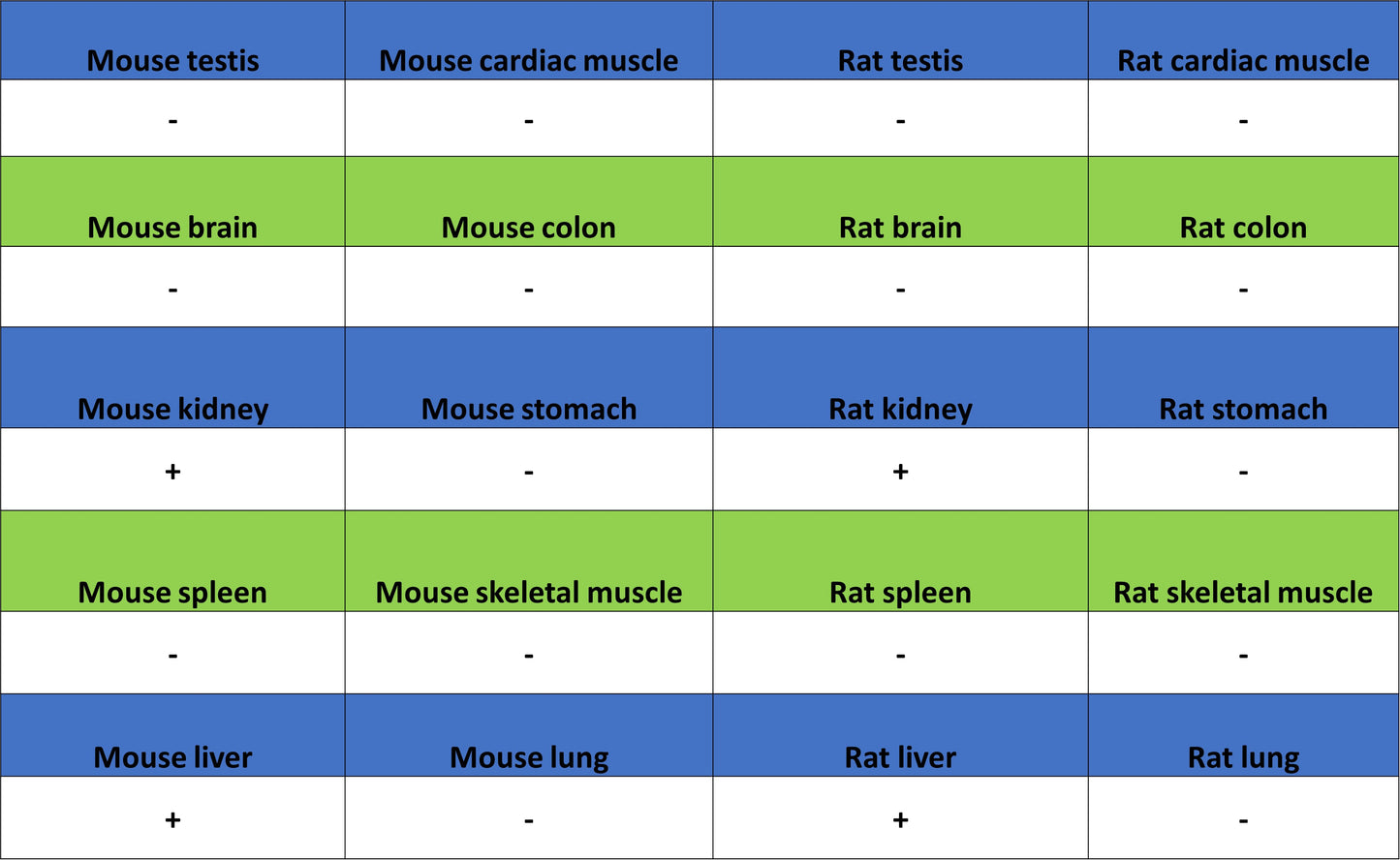
IHC shows positive staining in paraffin-embedded mouse kidney. Anti-Aass antibody was used at 1/500 dilution, followed by a HRP Polymer for Rabbit IgG (ready to use). Counterstained with hematoxylin. Heat mediated antigen retrieval with Tris/EDTA buffer pH9.0 was performed before commencing with IHC staining protocol.
IHC shows positive staining in paraffin-embedded rat kidney. Anti-Aass antibody was used at 1/500 dilution, followed by a HRP Polymer for Rabbit IgG (ready to use). Counterstained with hematoxylin. Heat mediated antigen retrieval with Tris/EDTA buffer pH9.0 was performed before commencing with IHC staining protocol.
IHC shows positive staining in paraffin-embedded rat liver. Anti-Aass antibody was used at 1/500 dilution, followed by a HRP Polymer for Rabbit IgG (ready to use). Counterstained with hematoxylin. Heat mediated antigen retrieval with Tris/EDTA buffer pH9.0 was performed before commencing with IHC staining protocol.