Unione® TR-FRET Human IL17A/IL17RA Binding Kit
Unione® TR-FRET Human IL17A/IL17RA Binding Kit

Product Details
Product Details
Product Specification
| Host | Human |
| Stability & Storage | -80℃ |
Background
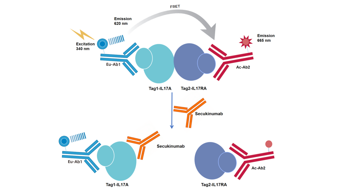
The kit employs homogeneous time-resolved fluorescence technology.TR-FRET), used for the determination ofIL17AwithIL17RAthe interactions between them. This method enables simple and rapid high-throughput identification of inhibitors and antibody blockers.
Components
Component |
Concentration |
100T |
500T |
2500T |
10000T |
Storage Temperature |
Tag1- IL17A protein |
100× |
5μL |
20μL |
100μL |
400μL |
-80℃ |
Tag2- IL17 RA protein |
100× |
5μL |
20μL |
100μL |
400μL |
-80℃ |
Anti-Tag1 Eu antibody |
50× |
10μL |
50μL |
250μL |
1000μL |
-80℃ |
Anti-Tag2 Ac antibody |
12.5× |
40μL |
200μL |
1000μL |
4000μL |
-80℃ |
Detection buffer |
10× |
400μL |
2mL |
10mL |
40mL |
-80℃ |
Caution: After the first thaw, immediately aliquot and store at the recommended temperature. Avoid storing diluted samples or subjecting them to repeated freeze-thaw cycles.
Protocol
1,Reagent preparation
1.1Before use, thaw all reagents at room temperature (allow to equilibrate at room temperature for at least 30 minutes). The reaction volume in a 384-well plate is 20 µL.muL (the reagent volumes for the reaction system are shown in the table). Calculate the required volume before preparation and prepare accordingly. The following formulation is for reference only, using 500 reactions as an example.
Table 1.Reagent preparation
Reagent name |
Configuration |
μL per test well |
Detection buffer |
Take 210 mLtimes Detection Add 18 mL of deionized water to the buffer, dilute.Explanationto1×, mixedEvenly prepareduse. |
- |
Sample to be tested |
According to the reaction system, the sample to be tested is diluted with 1× Detection buffer to the desired concentration..If DMSO is used, ensure that the DMSO concentration is consistent across all assay wells. |
2 |
Tag1- IL17A protein |
take20 μL Tag1-IL17A proteinUndiluted solution,1× Detection BufferDilute to2 mL,Mix well and set aside. |
4 |
Tag2- IL17 RA protein |
take20 μL Tag2-IL17 RA proteinUndiluted solution,1× Detection BufferDilute to2 mL,Mix well and set aside. |
4 |
Antibody Mix |
take50 μL Anti-Tag1 Eu antibodyConcentrate, add1× Detection buffer 2.45 mL,Mix well and set aside.; take200 μL Anti-Tag2 Ac antibodyConcentrate, add1× Detection buffer 2.3 mL,Mix well and set aside.; Anti-Tag1 Eu antibodyandAnti-Tag2 Ac antibodyAfter separate dilutions1:1Mix well and set aside. |
10 |
1.2Sample to be testedGradient dilution
withSecukinumabFor example, the diluent is 1× Detection buffer.To minimize the impact of matrix effects, it is recommended to dilute the sample with a solution that has the same matrix as the analyte; andThe sample to be tested should be adjusted according to its actual concentration.
Table2.Positive drug SsecukinumabGradient dilution(Adjust according to actual conditions)
SecukinumabFinal concentrationnM |
SecukinumabPreparation concentrationnM |
Preparation method |
|
① |
100 |
1000 |
1μL100μMSsecukinumab+99μL 1× Detection buffer |
② |
33.33 |
333.33 |
20μL ①+40μL 1× Detection buffer |
③ |
11.11 |
111.11 |
20μL ②+40μL 1× Detection buffer |
④ |
3.70 |
37.04 |
20μL ③+40μL 1× Detection buffer |
⑤ |
1.23 |
12.35 |
20μL ④+40μL 1× Detection buffer |
⑥ |
0.41 |
4.12 |
20μL ⑤+40μL 1× Detection buffer |
⑦ |
0.14 |
1.37 |
20muL ⑥+40muL 1× Detection buffer |
⑧ |
0.05 |
0.46 |
20muL ⑦+40muL 1× Detection buffer |
⑨ |
0.015 |
0.15 |
20muL ⑧+40muL 1× Detection buffer |
Blank |
0 |
0 |
40muL 1× Detection buffer |
2,Sample addition and controls
2.1Sample addition order for the experimental wells:2 μLSample to be tested,4 μL Tag1-IL17A proteinWorking fluid,4 μL Tag2-IL17 RA proteinWorking fluid,10 μL Antibody Mix, add in sequence384In the shallow-hole plate;
2.2Maximum value reference hole:2 μL 1× Detection buffer,4 μL Tag1-IL17A proteinWorking fluid,4 μL Tag2-IL17 RA proteinWorking fluid,10 μL Antibody Mix, add in sequence384In the shallow-hole plate;
2.3 Quality control well(NC):10Add μL of 1× Detection buffer.10μL AntibodyMix.
After all samples have been added, centrifuge the plate, seal it with a plate seal, and incubate at room temperature for 2 hours.
Maximum valueComparison |
sample |
NC |
|
Step one |
2 μL 1× DetectionBuffer |
2muL Gradient dilution of the sample to be tested |
10uL 1× DetectionBuffer |
4 μL Tag1-IL17A proteinWorking fluid | |||
Incubate for 10 minutes. | |||
|
Step two |
4 μLTag2- IL17 RA proteinnWorking fluid |
||
10μLMix | |||
Step three |
Cover with a sealing film and incubate at room temperature for 2 hours. |
||
3.Detection
Detect on a TR-FRET-compatible microplate reader..The excitation light is 320/340 nm, and detection is performed at 620 nm and 665 nm.postWavelength of the radiation.
[Result Calculation]
1)Calculate signal value(Ratio)The 665 nm fluorescence signal is multiplied by 10,000 compared to the 620 nm fluorescence signal..
Ratio = (665/620)times10000
2)Calculate based on the signal value.Net signal:
Net signal= (Std-NC)/NC×100
3)Calculate CV (%):
CV (%) = Standard Deviation/Mean Ratio × 100%
[Data Example]
The following data should not be used as a substitute for the data obtained in the experiment; it is provided for illustrative purposes only, and the results mayDue to the reading boardInstrumentIt varies from device to device.
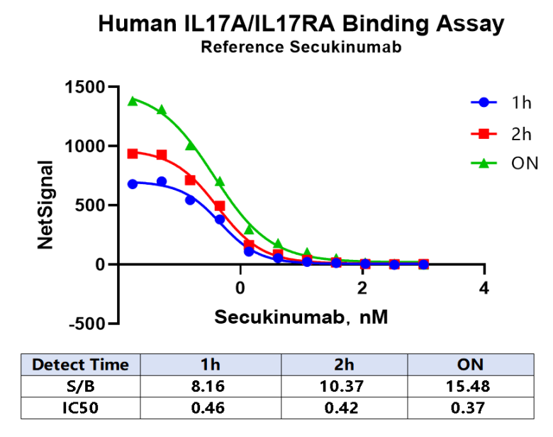
Note: RecommendedMicroplate (384-well plate, white, flat-bottom)