UniOne® TR-FRET Human CRBN Ligand Screening Assay Kit
UniOne® TR-FRET Human CRBN Ligand Screening Assay Kit

Product Details
Product Details
Product Specification
| Host | Human |
| Stability & Storage | -80℃ |
Background
PersonCereblon (CRBN) proteinLigandThe binding assay kit uses homogeneous time-resolved fluorescence technology (TR-FRET), which can be used for the detection and screening of CRBN inhibitors or ligands. It is a high-throughput screening method that enables rapid testing of CRBN inhibitors or ligands, with the basic principle shown in the figure below.
EuLabeled assay reagent (Anti-Tag1-Eu) specifically recognizes and binds toTag1-CRBN/DDB1protein, while the receptor-labeled thalidomide (Thalidomide-Ac) combines withCRBN, bringingEuandAcinto close proximity. Under external light excitation, energy resonance transfer occurs between the donor and acceptor, and by detecting the signal intensity at665nmwavelength, one can determinethe binding ofCRBN/DDB1withThalidomide-Ac.In the reaction system, the addedCRBNligand orPROTACcompetitively binds toCEBN/DDB1,causing the signal to decrease, and the ligand orPROTACAs the concentration increases, the signal decreases. This homogeneous assay is easy to perform and requires no washing steps.

Components
Component |
Concentration |
100T |
500T |
2500T |
10000T |
Storage Temperature |
Thalidomide |
10 mM in DMSO |
5μL |
5μL |
25μL |
100μL |
-80℃ |
Tag1-CRBN/DDB1 |
100× |
5μL |
25μL |
125μL |
500μL |
-80℃ |
Thalidomide-Ac |
100× |
5μL |
25μL |
125μL |
500μL |
-80℃ |
Anti-Tag1-Eu |
50× |
10μL |
50μL |
250μL |
1000μL |
-80℃ |
Detection buffer |
10× |
400μL |
2mL |
10mL |
40mL |
-80℃ |
Note: Allafter first thawingimmediatelydivide into aliquotsandstoreat the recommended storage temperature,Avoid storing after dilution and repeated freeze-thaw cycles..
Protocol
1,Reagent Preparation
1.1Before use, allow all reagents to thaw at room temperature (equilibrate at room temperature for at least 30 minutes). The reaction volume in a 384‑well plate is 20 µL.muL (reagent volumes for the reaction system are shown in the table). Calculate the required reaction volume before preparation and prepare accordingly. The following formulation is for reference only, using 500 reactions as an example.
Table 1.Reagent preparation
Reagent name |
Configuration |
μL per test well |
Detection buffer |
take2 mL 10timesDetection bufferJoin18 mL deionized water, diluteExplanationTo1×, mixedEvenly preparedUse. |
- |
Standard substance/Sample to be testedDilution |
Use diluted to1timesPrepare the samples to be tested and the standards using the assay buffer. If the stock solution of the sample is…DMSO, it is recommended to maintain the system withinDMSOThe content is consistent, and the maximum does not exceed2%.Standard dilutions are shown in the table below. |
5 |
Tag1-CRBN/DDB1 |
take25 μL of Tag1-CRBN/DDB1 stock solution, diluted with 1× Detection Buffer to a final volume of 2.5 mL,Mix thoroughly and set aside. |
5 |
Thalidomide-Ac |
take25 μL of thalidomide-ac stock solution, diluted with 1× detection buffer to a final volume of 2.5 mL,Mix thoroughly and set aside. |
5 |
Anti-Tag1-Eu |
take50 μL Anti-Tag1 EuOriginal solution, add1× Detection buffer 2.45 mL, Mix thoroughly and set aside. |
5 |
1.2Standard Reference Material Preparation Table
In the reagent kitThalidomideThe standard is10 mM DMSOMother solution. Please prepare the total amount of standard according to the experimental conditions; the following information is for reference only.
Use diluted to1timesPrepare the samples to be tested and the standards using the assay buffer. If the stock solution of the sample is…DMSO, it is recommended to maintain the system withinDMSOConsistent content,(The detection buffer does not containDMSO, please add according to the dilution of the standard or sample to be tested into1×detection bufferDMSO)and the maximum concentration should not exceed2%.
Table2.Standard Preparation Table
ThalidomidePreparation Concentration (nM) |
ThalidomideConcentration (nM) |
Preparation Method |
|
1 |
120000 |
30000 |
1 μL 10mMstock solution+ 82.3 μL 1× Detection buffer |
2 |
40000 |
10000 |
10 μL①+ 20 μL 1× Detection buffer |
3 |
13333.3 |
3333.33 |
10 μL②+ 20 μL 1× Detection buffer |
4 |
4444.4 |
1111.11 |
10 μL③+ 20 μL 1× Detection buffer |
5 |
1481.5 |
370.37 |
10 μL④+20 μL 1× Detection buffer |
6 |
493.8 |
123.46 |
10 μL⑤+20 μL 1× Detection buffer |
7 |
164.6 |
41.15 |
10μL⑥+20μL 1× Detection buffer |
8 |
54.9 |
13.72 |
10μL⑦+20μL 1× Detection buffer |
9 |
18.3 |
4.57 |
10μL⑧+20μL 1× Detection buffer |
10 |
6.1 |
1.52 |
10μL⑨+20μL 1× Detection buffer |
Blank |
0 |
0 |
20μL 1× Detection buffer |
Positive drug standard curve |
Sample to be tested |
Negative control well (NC) |
Eu control well |
|
Step one |
5μL Gradient dilutionThalidomide |
5μL Gradient dilution of the sample to be tested |
5μL 1× Detection buffer |
15μL 1×Detection buffer Add5μL 1×Anti-Tag1 Eu antibody |
5 μL Tag1-CRBN/DDB1 | ||||
|
Seal the plate,1000 rpm, centrifugefor 1 minutemix well, incubate at room temperaturefor 10 minutes | ||||
Step two |
10μL Thalidomide-Ac + Anti-Tag1-Eu 1:1 Mix |
|||
|
Seal the plate,1000 rpm, centrifugefor 1 minutemix well, incubate at room temperaturefor 120 minutes, read the plate | ||||
2andSample addition and controls
2.1Samples to be tested:5μL gradient dilutionsamples to be tested,5μLTag1-CRBN/DDB1working solution,5μLThalidomide-Acworking solution,5μLAnti-Tag1-Euworking solution (Thalidomide-Accan be mixed withAnti-Tag1-Euin proportionAfter mixing, add it sequentially to a 384-well plate.
2.2 Positive drug standard curve:5μL gradient dilutionThalidomide,5μLTag1-CRBN/DDB1Working fluid,5 μL Thalidomide-AcWorking fluid,5μLAnti-Tag1-EuWorking fluid.
2.3NegativeComparisonHole (NC):5μL 1× Detection buffer insteadTo be testedSample;
2.4Eu control hole: 15Add μL of 1× Detection buffer5μL of diluted Anti-Tag1 Eu antibody.
After adding all samples, centrifuge, seal with a plate cover, and incubate at room temperature for 2 hours.
3.Detection
Detection on a plate reader compatible with TR-FRET.The excitation wavelength is 320/340 nm, with detection at 620 nm and 665 nm.sendWavelength of the radiation.
[Result Calculation]
1)Calculate the signal value(Ratio): Multiply the 665nm fluorescence signal by the 620nm fluorescence signal, then multiply by 10000.
Ratio = (665/620)×10000
2Calculate CV (%):
CV (%) = Standard Deviation / Mean Ratio × 100%
[Data Example]
The following data cannot replace the data obtained in experiments; it is only for illustration, and results mayvary depending on the plate readerandinstrument.
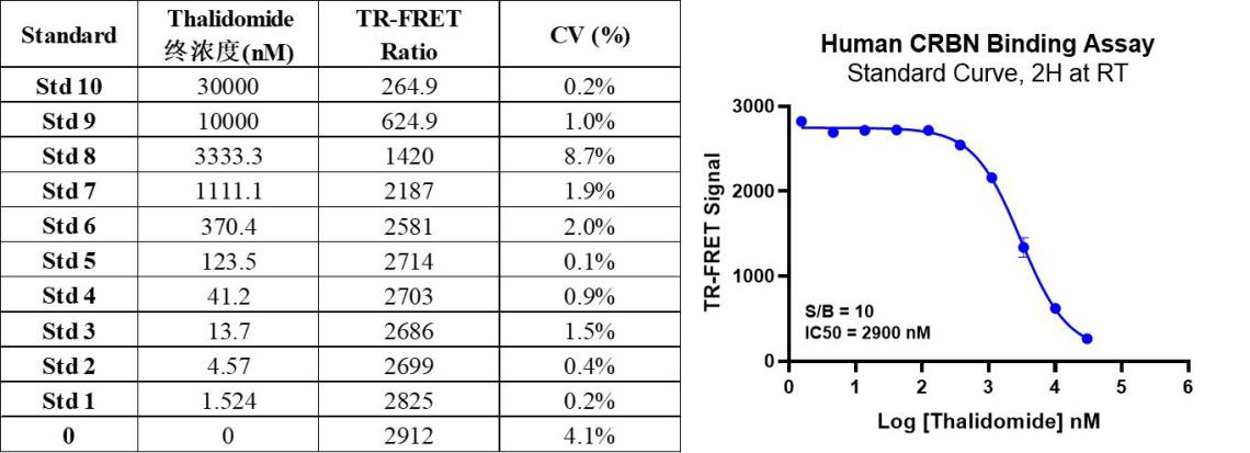
(1) Standard Curve*
As shown in the chart, due tothalidomidestandards competing withthalidomide-Acfor binding toCRBN/DDB1, the signal value decreases.
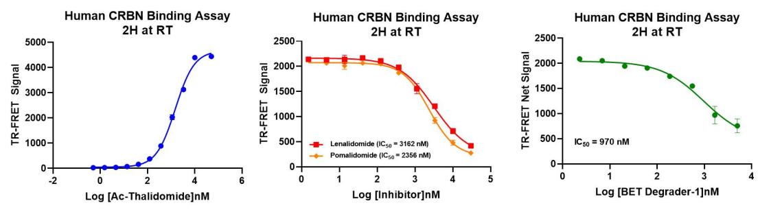
(2) Binding activity and inhibitor validation*
Thalidomide-Ac binding to CRBN/DDB1 was verified. The results show that when the protein concentration remains constant, the detection signal increases with increasing concentrations of Thalidomide-Ac. Lenalidomide, Pomalidomide, and PROTAC BET Degrader-1 can compete with Thalidomide-Ac for binding to CRBN/DDB1.
Note:*Data are for illustrative purposes only; recommendedMicroplate (384-well plate, white, shallow wells)