Standard curve
Example of IL-1β,IL-2,IL-4,IL-5,IL-6,IL-10,IL-12p70,TNF-α,IFN-γ ,CXCL1/KC standard curve in Assay Diluent .
Mouse Inflammation 10-Plex Kit (Flow Cytometry Multiplex Bead Assay)
Mouse Inflammation 10-Plex Kit (Flow Cytometry Multiplex Bead Assay)
Price:
Regular price
$3,000 USD
Regular price
Sale price
$3,000 USD
Unit price
per
For shipping services or bulk orders, you may request a quotation.
Secure checkout with

View full details
Product Details
Product Details
Product Specification
| Antigen | IL-1β、IL-2、IL-4、IL-5、IL-6、IL-10、IL-12p70、TNF-α、IFN-γ、CXCL1/KC |
| Reactivity | Mouse |
| Stability & Storage | 12 months from date of receipt / reconstitution, 2 to 8°C as supplied. |
Kit
| Precision | Intra-assay: <5%; Inter-assay: <15% |
| Sample type | Serum; Plasma; Cell culture supernatant |
| Assay type | Sandwich (quantitative) |
| Sensitivity | <3.0 pg/mL |
| Recovery | 70% - 130% |
| Assay time | 70 minutes |
| Species reactivity | Mouse |
Background
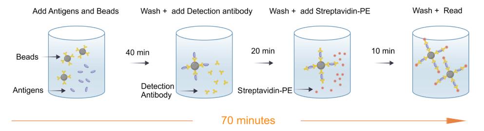
The Cytometric Bead Array (CBA) technology enables the capture of soluble analytes or analyte panels using beads with known sizes and fluorescence intensities, thereby allowing the detection of analytes via flow cytometry.
Each capture bead in the CBA kit is conjugated with a specific antibody. The detection reagent provided with the kit is Streptavidin-Phycoerythrin, and the intensity of the fluorescent signal generated by this reagent is proportional to the amount of bound analyte.
When the capture beads and detection reagent are incubated together with the test sample containing target analytes, sandwich complexes are formed. Detection of these complexes by flow cytometry allows for the identification of particles exhibiting the fluorescence characteristics of both the beads and the detection reagent.
Picture
Picture
CBA
Linearity
After serial dilution of the high-concentration combined proteins of mouse IL-1β,IL-2, IL-4, IL-5, IL-6, IL-10, IL-12p70,TNF-α,IFN-γ,CXCL1/KC using C57BL/6 mouse serum matrices, the concentrations of mouse IL-1β,IL-2, IL-4, IL-5, IL-6, IL-10, IL-12p70,TNF-α,IFN-γ,CXCL1/KC were measured and interpolated from the target standard curves and corrected for sample dilution.
Linearity
The high-concentration combined protein was serially diluted using the EDTA plasma matrix of C57BL/6 mice.
Linearity
The high-concentration combined protein was serially diluted using the heparin plasma matrix of C57BL/6 mice.
Linearity
The high-concentration combined protein was serially diluted using the citrate plasma matrix of C57BL/6 mice.
Linearity
The high-concentration combined protein was serially diluted using the serum matrix of BALB/c mice.
Linearity
The high-concentration combined protein was serially diluted using the EDTA plasma matrix of BALB/c mice.
Linearity
The high-concentration combined protein was serially diluted using the heparin plasma matrix of BALB/c mice.
Linearity
The high-concentration combined protein was serially diluted using the citrate plasma matrix of BALB/c mice.
Recovery
Within the detection range, the combined cytokine proteins were added to different matrices at high,medium and low concentrations.
The matrices used in these experiments were diluted 2-fold with the assay buffer before the addition of the cytokine proteins.
The results were compared with those of samples that had the same concentration of cytokines added in the assay buffer.
Recovery
Within the detection range, the combined cytokine proteins were added to different matrices at high,medium and low concentrations.
The matrices used in these experiments were diluted 2-fold with the assay buffer before the addition of the cytokine proteins.
The results were compared with those of samples that had the same concentration of cytokines added in the assay buffer.
Recovery
Within the detection range, the combined cytokine proteins were added to different matrices at high,medium and low concentrations.
The matrices used in these experiments were diluted 2-fold with the assay buffer before the addition of the cytokine proteins.
The results were compared with those of samples that had the same concentration of cytokines added in the assay buffer.
Recovery
Within the detection range, the combined cytokine proteins were added to different matrices at high,medium and low concentrations.
The matrices used in these experiments were diluted 2-fold with the assay buffer before the addition of the cytokine proteins.
The results were compared with those of samples that had the same concentration of cytokines added in the assay buffer.
Recovery
Within the detection range, the combined cytokine proteins were added to different matrices at high,medium and low concentrations.
The matrices used in these experiments were diluted 2-fold with the assay buffer before the addition of the cytokine proteins.
The results were compared with those of samples that had the same concentration of cytokines added in the assay buffer.
Quantifiction of Mouse spleen cells
Mouse spleen cells Unsimulated Supernatant: The cells were cultured for24 hours, the cell culture supernatant was collected and analyzed quantitatively using S0Q1005.
Mouse spleen cells simulated Supernatant: The cells of were stimulated with 10 μg/mL ConA. After 24 hours, the cell culture supernatant was collected and analyzed quantitatively using S0Q1005.
Intra-assay precision
Ten replicates of each of three different levels of IL-1β,IL-2, IL-4, IL-5, IL-6, IL-10, IL-12p70,TNF-α,IFN-γ and CXCL1/KC were tested.