M1
Mouse RAW264.7 Cell Polarization Induction Kit II
Mouse RAW264.7 Cell Polarization Induction Kit II
Price:
Regular price
$716 USD
Regular price
Sale price
$716 USD
Unit price
per
For shipping services or bulk orders, you may request a quotation.
Secure checkout with

View full details
Product Details
Product Details
Product Specification
| Species | Mouse | ||||
| Synonyms | Mouse RAW264.7-Cell Polarization Induction Kit | ||||
| Reconstitution |
|
||||
| Stability & Storage |
|
Components
Contain |
Formulation |
Reference dosage |
S Size |
M Size |
IFN-γ Protein, Mouse |
Lyophilized Powder |
25ng/ml |
65μg |
125μg |
IL-4 Protein, Mouse |
Lyophilized Powder |
20ng/ml |
50μg |
100μg |
Lipopolysaccharide (LPS) solution (500X, 2.5mg/ml) |
Liquid |
100ng/ml |
100μl |
100μl*2 |
Protocol
Experimental Cells: RAW264.7
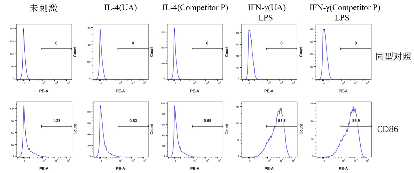
I. M1 Polarization
Culture Conditions: 6-well cell culture plate, 2 × 10⁵ cells per well (excessive cell density may impair polarization efficiency), incubated at 37°C with 5% CO₂ for 24 hours. Replace the medium with DMEM + 2% FBS (to reduce interference from cytokines in serum) and add the corresponding cytokines as indicated in the table below. Continue culturing at 37°C with 5% CO₂ for 24 hours.
| Category | Constituent | Final Conc (ng/ml) |
| M1 |
IFN-γ LPS |
25 100 |
| Negative control | Cell Only |
Flow Cytometry Detection Procedure:
Cell Collection: After 24 hours of cytokine treatment, discard the supernatant medium, wash the cells once with PBS, discard the supernatant, then gently resuspend adherent cells by pipetting with PBS. Centrifuge to collect cells.
Cell Counting: Resuspend cells in 0.5 mL of 1% BSA, calculate the total cell number, and assess cell viability, which should be ≥95%.
Antibody Incubation: Add PE Rat Anti-Mouse CD86 mAb (antibody dosage as per manufacturer’s instructions) or the corresponding isotype control. Incubate at room temperature for 30 minutes.
Cell Washing: Wash cells with PBS to remove residual antibodies, then resuspend in PBS again.
Viability Staining: Add Fixable Viability Kit(dosage as per manufacturer’s instructions) to each well. Incubate at room temperature for 15 minutes, protected from light.
Cell Washing: Wash cells with 1% BSA to remove residual antibodies, then resuspend in 1% BSA.
Flow Cytometry Analysis.
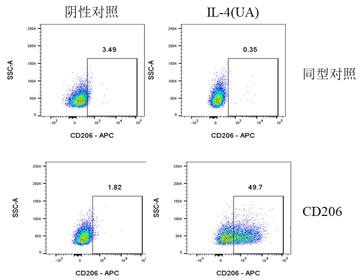
II. M2 Polarization
Culture Conditions: 6-well cell culture plate, 2 × 10⁵ cells per well (excessive cell density may impair polarization efficiency), cultured at 37°C with 5% CO₂ for 24 hours. Replace the medium with DMEM + 2% FBS (to reduce interference from cytokines in serum) and add the corresponding cytokines as indicated in the table below. Continue culturing at 37°C with 5% CO₂ for 24 hours.
| Category | Constituent | Final Conc (ng/ml) |
| M2 | IL-4 | 20 |
| Negative control | Cell only |
Flow Cytometry Detection Procedure:
Cell Collection: After 24 hours of cytokine treatment, discard the supernatant medium, wash the cells once with PBS, discard the supernatant, then gently resuspend adherent cells by pipetting with PBS. Centrifuge to collect cells.
Cell Counting: Resuspend cells in 0.5 mL of 1% BSA, calculate the total cell number, and assess cell viability, which should be ≥95%.
Viability Staining: After centrifugation, resuspend cells in PBS to a density of 1 × 10⁷ cells/mL. Add 100 µL per well/tube to a 96-well plate or flow cytometry tube, and add the Zombie Violet™ Fixable Viability Kit (dosage as per manufacturer's instructions). Incubate at room temperature, protected from light, for 15 minutes. Centrifuge at 300 × g for 5 minutes and discard the supernatant.
Fixation: Resuspend and fix cells in 0.2 mL of 4% Paraformaldehyde (PFA). Incubate at room temperature, protected from light, for 15 minutes. Centrifuge at 300 × g for 5 minutes and discard the supernatant (to remove residual fixative).
Permeabilization: Resuspend and permeabilize cells in 0.2 mL of 1X Permeabilization Buffer . Incubate at room temperature, protected from light, for 30 minutes. Centrifuge at 400 × g for 5 minutes and discard the supernatant.
Wash: Add 0.2 mL of 1X Permeabilization Buffer to resuspend cells. Centrifuge at 400 × g for 5 minutes and discard the supernatant.
Blocking: Add 0.1 mL of 1X Permeabilization Buffer to each well/tube, then add 2 µg of CD16/CD32 antibody (S0B7003) for blocking. Incubate on ice for 30 minutes. Centrifuge at 400 × g for 5 minutes and discard the supernatant.
Antibody Incubation: Add 0.1 mL of 1X Permeabilization Buffer to each well/tube, then add APC anti-Mouse CD206 (MMR) mAb (antibody dosage as per manufacturer's instructions) or the corresponding isotype control (APC Rat IgG Isotype Ctrl Antibody). Incubate at room temperature for 30 minutes. Centrifuge at 400 × g for 5 minutes and discard the supernatant (to remove residual antibody).
Cell Washing: Add 0.2 mL of 1% BSA to wash the cells. Centrifuge at 400 × g for 5 minutes and discard the supernatant. Repeat this wash step twice. Finally, resuspend the cells in 200 µL of 1% BSA.
Flow Cytometry Analysis.
Picture
Picture
FC
M2