WB result of SLC7A5 Recombinant Rabbit mAb
Primary antibody: SLC7A5 Recombinant Rabbit mAb at 1/1000 dilution
Lane 1: F9 whole cell lysate 20 µg
Secondary antibody: Goat Anti-Rabbit IgG (H+L), HRP conjugated at 1/10000 dilution
Predicted MW: 56 kDa
Observed MW: 38 kDa
SLC7A5 Recombinant Rabbit mAb (S-4496-55)
SLC7A5 Recombinant Rabbit mAb (S-4496-55)
Price:
Regular price
$100 USD
Regular price
Sale price
$100 USD
Unit price
per
For shipping services or bulk orders, you may request a quotation.
Secure checkout with

View full details
Product Details
Product Details
Product Specification
| Host | Rabbit |
| Antigen | SLC7A5 |
| Synonyms | Large neutral amino acids transporter small subunit 1; 4F2 light chain (4F2 LC; 4F2LC); L-type amino acid transporter 1 (LAT1); Solute carrier family 7 member 5; Lat1; Slc7a5 |
| Immunogen | Synthetic Peptide |
| Location | Lysosome, Cell membrane |
| Accession | Q9Z127 |
| Clone Number | S-4496-55 |
| Antibody Type | Recombinant mAb |
| Isotype | IgG |
| Application | WB, IHC-P, ICC, IF |
| Reactivity | Ms, Rt |
| Positive Sample | F9, mouse testis, mouse brain, mouse pancreas, rat testis, rat brain |
| Purification | Protein A |
| Concentration | 2 mg/ml |
| Conjugation | Unconjugated |
| Physical Appearance | Liquid |
| Storage Buffer | PBS, 40% Glycerol, 0.05% BSA, 0.02% sodium azide |
| Stability & Storage | 12 months from date of receipt / reconstitution, -20 °C as supplied |
Dilution
| application | dilution | species |
| WB | 1:1000 | Ms, Rt |
| IHC-P | 1:2000 | Ms, Rt |
| ICC | 1:400 | Ms |
| IF | 1:2000 | Ms, Rt |
Background
SLC7A5, also known as L-type amino acid transporter 1 (LAT1), is a key member of the solute carrier family 7 and plays a central role in cellular physiology and disease. Its critical function is to form a sodium-independent heterodimeric complex with SLC3A2, acting as a high-affinity transporter that mediates the uptake of large neutral amino acids such as leucine, phenylalanine, and tryptophan, and also participates in the transmembrane transport of thyroid hormones. Functionally, SLC7A5 not only serves as the primary channel for cells to acquire essential amino acids, but also activates the mTORC1 signaling pathway by regulating lysosomal leucine concentration, thereby functioning as a key nutrient sensor that controls cell growth and metabolism. Consequently, aberrant expression of SLC7A5 is closely associated with various pathological conditions: it is particularly prominent in cancer, being significantly upregulated in numerous tumors including breast cancer, colorectal cancer, and glioma, where it is tightly linked to poor patient prognosis and chemoresistance by promoting proliferation and metabolic reprogramming; simultaneously, it is also deeply involved in the activation of immune cells (such as T cells and B cells), modulating inflammatory processes. Given its dual importance in tumorigenesis and immune regulation, SLC7A5 has emerged as a highly promising diagnostic biomarker and precision therapeutic target in oncology.
Picture
Picture
Western Blot
WB result of SLC7A5 Recombinant Rabbit mAb
Primary antibody: SLC7A5 Recombinant Rabbit mAb at 1/1000 dilution
Lane 1: mouse liver lysate 20 µg
Lane 2: mouse testis lysate 20 µg
Lane 3: mouse brain lysate 20 µg
Lane 4: mouse pancreas lysate 20 µg
Negative control: mouse liver lysate 20 µg
Secondary antibody: Goat Anti-Rabbit IgG (H+L), HRP conjugated at 1/10000 dilution
Predicted MW: 56 kDa
Observed MW: 38 kDa
This blot was developed with high sensitivity substrate
WB result of SLC7A5 Recombinant Rabbit mAb
Primary antibody: SLC7A5 Recombinant Rabbit mAb at 1/1000 dilution
Lane 1: rat liver lysate 20 µg
Lane 2: rat testis lysate 20 µg
Lane 3: rat brain lysate 20 µg
Negative control: rat liver lysate 20 µg
Secondary antibody: Goat Anti-Rabbit IgG (H+L), HRP conjugated at 1/10000 dilution
Predicted MW: 56 kDa
Observed MW: 38 kDa
This blot was developed with high sensitivity substrate
Immunohistochemistry
IHC shows positive staining in paraffin-embedded mouse cerebral cortex. Anti-SLC7A5 antibody was used at 1/2000 dilution, followed by a HRP Polymer for Rabbit IgG (ready to use). Counterstained with hematoxylin. Heat mediated antigen retrieval with Tris/EDTA buffer pH9.0 was performed before commencing with IHC staining protocol.
IHC shows positive staining in paraffin-embedded mouse testis. Anti-SLC7A5 antibody was used at 1/2000 dilution, followed by a HRP Polymer for Rabbit IgG (ready to use). Counterstained with hematoxylin. Heat mediated antigen retrieval with Tris/EDTA buffer pH9.0 was performed before commencing with IHC staining protocol.
Negative control: IHC shows negative staining in paraffin-embedded mouse liver. Anti-SLC7A5 antibody was used at 1/2000 dilution, followed by a HRP Polymer for Rabbit IgG (ready to use). Counterstained with hematoxylin. Heat mediated antigen retrieval with Tris/EDTA buffer pH9.0 was performed before commencing with IHC staining protocol.
IHC shows positive staining in paraffin-embedded rat testis. Anti-SLC7A5 antibody was used at 1/2000 dilution, followed by a HRP Polymer for Rabbit IgG (ready to use). Counterstained with hematoxylin. Heat mediated antigen retrieval with Tris/EDTA buffer pH9.0 was performed before commencing with IHC staining protocol.
Negative control: IHC shows negative staining in paraffin-embedded rat liver. Anti-SLC7A5 antibody was used at 1/2000 dilution, followed by a HRP Polymer for Rabbit IgG (ready to use). Counterstained with hematoxylin. Heat mediated antigen retrieval with Tris/EDTA buffer pH9.0 was performed before commencing with IHC staining protocol.
Immunocytochemistry
ICC shows positive staining in F9 cells. Anti- SLC7A5 antibody was used at 1/400 dilution (Green) and incubated overnight at 4°C. Goat polyclonal Antibody to Rabbit IgG - H&L (Alexa Fluor® 488) was used as secondary antibody at 1/1000 dilution. The cells were fixed with 100% ice-cold methanol and permeabilized with 0.1% PBS-Triton X-100. Nuclei were counterstained with DAPI (Blue). Counterstain with tubulin (Red).
Immunofluorescence
IF shows positive staining in paraffin-embedded mouse testis. Anti-SLC7A5 antibody was used at 1/2000 dilution (Green) and incubated overnight at 4°C. Goat polyclonal Antibody to Rabbit IgG - H&L (Alexa Fluor® 488) was used as secondary antibody at 1/1000 dilution. Counterstained with DAPI (Blue). Heat mediated antigen retrieval with EDTA buffer pH9.0 was performed before commencing with IF staining protocol.
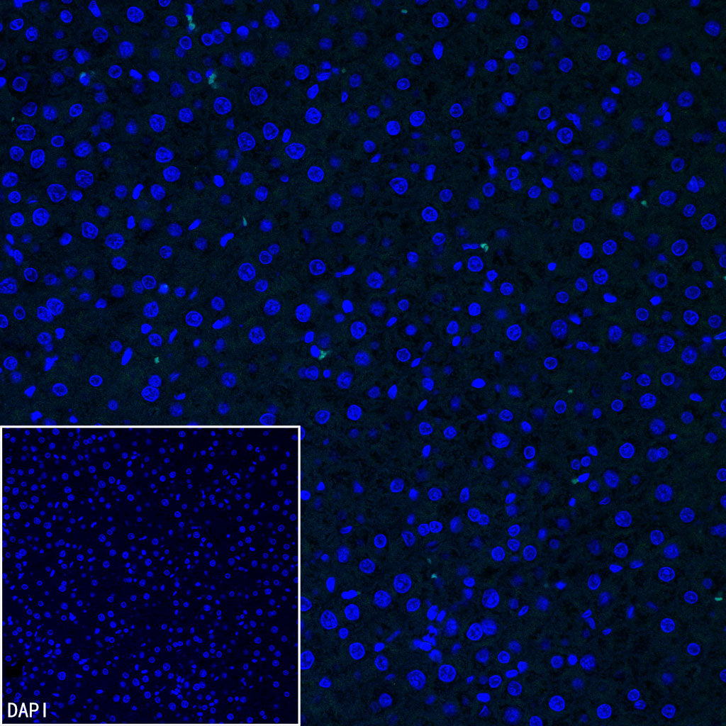
Negative control: IF shows negative staining in paraffin-embedded mouse liver. Anti-SLC7A5 antibody was used at 1/2000 dilution and incubated overnight at 4°C. Goat polyclonal Antibody to Rabbit IgG - H&L (Alexa Fluor® 488) was used as secondary antibody at 1/1000 dilution. Counterstained with DAPI (Blue). Heat mediated antigen retrieval with EDTA buffer pH9.0 was performed before commencing with IF staining protocol.
IF shows positive staining in paraffin-embedded rat testis. Anti-SLC7A5 antibody was used at 1/2000 dilution (Green) and incubated overnight at 4°C. Goat polyclonal Antibody to Rabbit IgG - H&L (Alexa Fluor® 488) was used as secondary antibody at 1/1000 dilution. Counterstained with DAPI (Blue). Heat mediated antigen retrieval with EDTA buffer pH9.0 was performed before commencing with IF staining protocol.
Negative control: IF shows negative staining in paraffin-embedded rat liver. Anti-SLC7A5 antibody was used at 1/2000 dilution and incubated overnight at 4°C. Goat polyclonal Antibody to Rabbit IgG - H&L (Alexa Fluor® 488) was used as secondary antibody at 1/1000 dilution. Counterstained with DAPI (Blue). Heat mediated antigen retrieval with EDTA buffer pH9.0 was performed before commencing with IF staining protocol.