WB result of RPL24 Rabbit pAb
Primary antibody: RPL24 Rabbit pAb at 1/1000 dilution
Lane 1: Raji whole cell lysate 20 µg
Lane 2: HL-60 whole cell lysate 20 µg
Lane 3: A431 whole cell lysate 20 µg
Lane 4: HT-29 whole cell lysate 20 µg
Lane 5: HT-1080 whole cell lysate 20 µg
Lane 6: HEK-293 whole cell lysate 20 µg
Lane 7: HeLa whole cell lysate 20 µg
Secondary antibody: Goat Anti- rabbit IgG, (H+L), HRP conjugated at 1/10000 dilution
Predicted MW: 18 kDa
Observed MW: 25 kDa
RPL24 Rabbit Polyclonal Antibody
RPL24 Rabbit Polyclonal Antibody
Price:
Regular price
$100 USD
Regular price
Sale price
$100 USD
Unit price
per
For shipping services or bulk orders, you may request a quotation.
Secure checkout with

View full details
Product Details
Product Details
Product Specification
| Host | Rabbit |
| Antigen | RPL24 |
| Synonyms | Large ribosomal subunit protein eL24; 60S ribosomal protein L24; 60S ribosomal protein L30 |
| Immunogen | Synthetic Peptide |
| Location | Cytoplasm |
| Accession | P83731 |
| Antibody Type | Polyclonal antibody |
| Isotype | IgG |
| Application | WB, ICC |
| Reactivity | Hu, Ms, Rt, Mk |
| Positive Sample | Raji, HL-60, A431, HT-29, HT-1080, HEK-293, HeLa, NIH/3T3, PC-12, COS-7 |
| Purification | Immunogen Affinity |
| Concentration | 0.5 mg/ml |
| Conjugation | Unconjugated |
| Physical Appearance | Liquid |
| Storage Buffer | PBS, 40% Glycerol, 0.05% BSA, 0.02% sodium azide |
| Stability & Storage | 12 months from date of receipt / reconstitution, -20 °C as supplied |
Dilution
| application | dilution | species |
| WB | 1:1000-1:2000 | Hu, Ms, Rt, Mk |
| ICC | 1:500 | Hu |
Background
RPL24, also known as ribosomal protein L24, is a key component of the eukaryotic 60S large ribosomal subunit and belongs to the ribosomal protein family. This protein is localized in the nucleus and plays a dual role in ribosome biogenesis: it not only participates as a structural protein in the folding and stabilization of ribosomal RNA (rRNA), but also interacts with the pre-rRNA processing complex in the nucleolus to promote the maturation of 28S rRNA. At the functional level, RPL24 protein is essential for maintaining ribosomal integrity and translational efficiency, and alterations in its expression levels directly affect the protein synthesis capacity of cells. Beyond its classical ribosomal function, recent studies have also revealed extraribosomal functions of RPL24 protein, such as binding to MDM2 protein and inhibiting its activity, thereby regulating cell cycle and apoptosis through the p53 signaling pathway, while also directly binding to the p53 tumor suppressor protein itself and enhancing its transcriptional activity. Mutations or aberrant expression of the RPL24 gene are closely associated with various diseases; for example, upregulation of RPL24 has been observed in multiple cancers, where its overexpression may drive tumor initiation and progression by promoting the translation of pro-oncogenic proteins, while haploinsufficiency of the RPL24 gene has been confirmed to be associated with congenital erythroid hypoplasia in certain hereditary ribosomopathies such as Diamond-Blackfan anemia. Therefore, RPL24 protein is not only a classical ribosomal component but also a signaling hub that establishes an important link between ribosomal function and tumor suppressor regulation, making it a highly attractive molecule in cancer and ribosome-related disease research.
Picture
Picture
Western Blot
WB result of RPL24 Rabbit pAb
Primary antibody: RPL24 Rabbit pAb at 1/1000 dilution
Lane 1: NIH/3T3 whole cell lysate 20 µg
Secondary antibody: Goat Anti- rabbit IgG, (H+L), HRP conjugated at 1/10000 dilution
Predicted MW: 18 kDa
Observed MW: 25 kDa
WB result of RPL24 Rabbit pAb
Primary antibody: RPL24 Rabbit pAb at 1/1000 dilution
Lane 1: PC-12 whole cell lysate 20 µg
Secondary antibody: Goat Anti- rabbit IgG, (H+L), HRP conjugated at 1/10000 dilution
Predicted MW: 18 kDa
Observed MW: 25 kDa
WB result of RPL24 Rabbit pAb
Primary antibody: RPL24 Rabbit pAb at 1/1000 dilution
Lane 1: COS-7 whole cell lysate 20 µg
Secondary antibody: Goat Anti- rabbit IgG, (H+L), HRP conjugated at 1/10000 dilution
Predicted MW: 18 kDa
Observed MW: 25 kDa
Immunocytochemistry
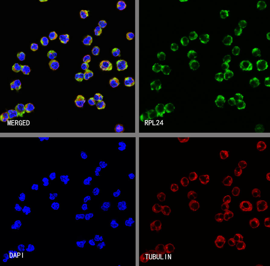
ICC shows positive staining in Raji cells. Anti- RPL24 antibody was used at 1/500 dilution (Green) and incubated overnight at 4°C. Goat polyclonal Antibody to Rabbit IgG - H&L (Alexa Fluor® 488) was used as secondary antibody at 1/1000 dilution. The cells were fixed with 4% PFA and permeabilized with 0.1% PBS-Triton X-100. Nuclei were counterstained with DAPI (Blue). Counterstain with tubulin (Red).