WB result of PTK7 Recombinant Rabbit mAb
Primary antibody: PTK7 Recombinant Rabbit mAb at 1/1000 dilution
Lane 1: HL-60 whole cell lysate 20 μg
Lane 2: HCT-116 whole cell lysate 20 μg
Lane 3: SK-OV-3 whole cell lysate 20 μg
Lane 4: PANC-1 whole cell lysate 20 μg
Lane 5: A204 whole cell lysate 20 μg
Negative control: HL-60 whole cell lysate
Secondary antibody: Goat Anti-Rabbit IgG (H+L), HRP conjugated at 1/10000 dilution
Predicted MW: 118 kDa
Observed MW: 118-140 kDa
PTK7 Recombinant Rabbit mAb ,PBS Only(SDT-2432-156)
PTK7 Recombinant Rabbit mAb ,PBS Only(SDT-2432-156)
Price:
Regular price
$920 USD
Regular price
Sale price
$920 USD
Unit price
per
For shipping services or bulk orders, you may request a quotation.
Secure checkout with

View full details
Product Details
Product Details
Product Specification
| Host | Rabbit |
| Antigen | PTK7 |
| Synonyms | Inactive tyrosine-protein kinase 7; Colon carcinoma kinase 4 (CCK-4); Protein-tyrosine kinase 7; Pseudo tyrosine kinase receptor 7; Tyrosine-protein kinase-like 7; CCK4 |
| Immunogen | Recombinant Protein |
| Location | Cell membrane |
| Accession | Q13308 |
| Clone Number | SDT-2432-156 |
| Antibody Type | Recombinant mAb |
| Isotype | IgG |
| Application | WB, IHC-P |
| Reactivity | Hu, Ms, Rt, Mk |
| Positive Sample | HCT-116, SK-OV-3, PANC-1, A204, C2C12, PC-12, COS7 |
| Purification | Protein A |
| Concentration | 1 mg/ml |
| Conjugation | Unconjugated |
| Physical Appearance | Liquid |
| Storage Buffer | PBS |
| Stability & Storage | 12 months from date of receipt / reconstitution, 4 °C as supplied |
Dilution
| application | dilution | species |
| WB | 1:1000-1:5000 | Hu, Ms, Rt, Mk |
| IHC-P | 1:2000 | Hu, Ms, Rt |
Background
PTK7 (Protein Tyrosine Kinase 7), also known as CCK-4, is a pseudokinase belonging to the receptor tyrosine kinase (RTK) family. Although it possesses a typical extracellular domain, transmembrane region, and intracellular kinase domain, it lacks intrinsic kinase activity due to mutations in several key catalytic residues within its intracellular kinase domain, and thus mainly relies on interactions with other proteins to mediate signal transduction. During embryonic development, PTK7 serves as an important regulator of the non-canonical Wnt signaling pathway, modulating cell polarity and directional migration through its involvement in the planar cell polarity (PCP) pathway, which is essential for critical morphogenetic processes such as neural tube closure, cardiac development, and limb formation. Furthermore, PTK7 is expressed at low levels in most adult tissues but is significantly overexpressed in various malignancies, including colon cancer, lung cancer, breast cancer, and acute myeloid leukemia, where its overexpression is closely associated with tumor invasion, metastasis, and poor prognosis, making it a promising tumor biomarker and drug target. Currently, antibody-drug conjugates targeting PTK7, such as Cofetuzumab pelidotin, have entered clinical trials to explore their potential in cancer targeted therapy.
Picture
Picture
Western Blot
WB result of PTK7 Recombinant Rabbit mAb
Primary antibody: PTK7 Recombinant Rabbit mAb at 1/1000 dilution
Lane 1: C2C12 whole cell lysate 20 μg
Secondary antibody: Goat Anti-Rabbit IgG (H+L), HRP conjugated at 1/10000 dilution
Predicted MW: 118 kDa
Observed MW: 118-140 kDa
WB result of PTK7 Recombinant Rabbit mAb
Primary antibody: PTK7 Recombinant Rabbit mAb at 1/1000 dilution
Lane 1: PC-12 whole cell lysate 20 μg
Secondary antibody: Goat Anti-Rabbit IgG (H+L), HRP conjugated at 1/10000 dilution
Predicted MW: 118 kDa
Observed MW: 118-140 kDa
WB result of PTK7 Recombinant Rabbit mAb
Primary antibody: PTK7 Recombinant Rabbit mAb at 1/1000 dilution
Lane 1: COS7 whole cell lysate 20 μg
Secondary antibody: Goat Anti-Rabbit IgG (H+L), HRP conjugated at 1/10000 dilution
Predicted MW: 118 kDa
Observed MW: 118-140 kDa
Immunohistochemistry
IHC shows positive staining in paraffin-embedded human colon. Anti-PTK7 antibody was used at 1/2000 dilution, followed by a HRP Polymer for Rabbit IgG (ready to use). Counterstained with hematoxylin. Heat mediated antigen retrieval with Tris/EDTA buffer pH9.0 was performed before commencing with IHC staining protocol.
IHC shows positive staining in paraffin-embedded human stomach. Anti-PTK7 antibody was used at 1/2000 dilution, followed by a HRP Polymer for Rabbit IgG (ready to use). Counterstained with hematoxylin. Heat mediated antigen retrieval with Tris/EDTA buffer pH9.0 was performed before commencing with IHC staining protocol.
IHC shows positive staining in paraffin-embedded human breast cancer. Anti-PTK7 antibody was used at 1/2000 dilution, followed by a HRP Polymer for Rabbit IgG (ready to use). Counterstained with hematoxylin. Heat mediated antigen retrieval with Tris/EDTA buffer pH9.0 was performed before commencing with IHC staining protocol.
IHC shows positive staining in paraffin-embedded human colon cancer. Anti-PTK7 antibody was used at 1/2000 dilution, followed by a HRP Polymer for Rabbit IgG (ready to use). Counterstained with hematoxylin. Heat mediated antigen retrieval with Tris/EDTA buffer pH9.0 was performed before commencing with IHC staining protocol.
IHC shows positive staining in paraffin-embedded human liver cancer. Anti-PTK7 antibody was used at 1/2000 dilution, followed by a HRP Polymer for Rabbit IgG (ready to use). Counterstained with hematoxylin. Heat mediated antigen retrieval with Tris/EDTA buffer pH9.0 was performed before commencing with IHC staining protocol.
IHC shows positive staining in paraffin-embedded human lung cancer. Anti-PTK7 antibody was used at 1/2000 dilution, followed by a HRP Polymer for Rabbit IgG (ready to use). Counterstained with hematoxylin. Heat mediated antigen retrieval with Tris/EDTA buffer pH9.0 was performed before commencing with IHC staining protocol.
Negative control: IHC shows negative staining in paraffin-embedded human cardiac muscle. Anti-PTK7 antibody was used at 1/2000 dilution, followed by a HRP Polymer for Rabbit IgG (ready to use). Counterstained with hematoxylin. Heat mediated antigen retrieval with Tris/EDTA buffer pH9.0 was performed before commencing with IHC staining protocol.
IHC shows positive staining in paraffin-embedded HCT 116 cell pellet. Anti-PTK7 antibody was used at 1/2000 dilution, followed by a HRP Polymer for Rabbit IgG (ready to use). Counterstained with hematoxylin. Heat mediated antigen retrieval with Tris/EDTA buffer pH9.0 was performed before commencing with IHC staining protocol.
IHC shows positive staining in paraffin-embedded SK-OV-3 cell pellet. Anti-PTK7 antibody was used at 1/2000 dilution, followed by a HRP Polymer for Rabbit IgG (ready to use). Counterstained with hematoxylin. Heat mediated antigen retrieval with Tris/EDTA buffer pH9.0 was performed before commencing with IHC staining protocol.
Negative control: IHC shows negative staining in paraffin-embedded human HL-60 cell pellet. Anti-PTK7 antibody was used at 1/2000 dilution, followed by a HRP Polymer for Rabbit IgG (ready to use). Counterstained with hematoxylin. Heat mediated antigen retrieval with Tris/EDTA buffer pH9.0 was performed before commencing with IHC staining protocol.
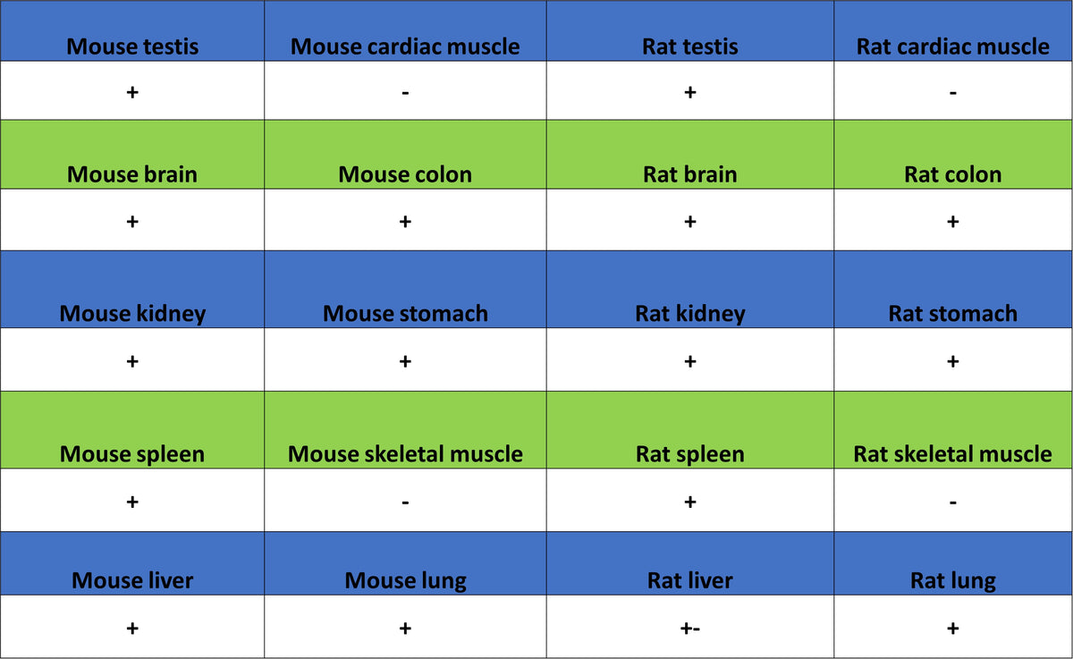
IHC shows positive staining in paraffin-embedded mouse colon. Anti-PTK7 antibody was used at 1/2000 dilution, followed by a HRP Polymer for Rabbit IgG (ready to use). Counterstained with hematoxylin. Heat mediated antigen retrieval with Tris/EDTA buffer pH9.0 was performed before commencing with IHC staining protocol.
Negative control: IHC shows negative staining in paraffin-embedded mouse skeletal muscle. Anti-PTK7 antibody was used at 1/2000 dilution, followed by a HRP Polymer for Rabbit IgG (ready to use). Counterstained with hematoxylin. Heat mediated antigen retrieval with Tris/EDTA buffer pH9.0 was performed before commencing with IHC staining protocol.
IHC shows positive staining in paraffin-embedded rat colon. Anti-PTK7 antibody was used at 1/2000 dilution, followed by a HRP Polymer for Rabbit IgG (ready to use). Counterstained with hematoxylin. Heat mediated antigen retrieval with Tris/EDTA buffer pH9.0 was performed before commencing with IHC staining protocol.
Negative control: IHC shows negative staining in paraffin-embedded rat cardiac muscle. Anti-PTK7 antibody was used at 1/2000 dilution, followed by a HRP Polymer for Rabbit IgG (ready to use). Counterstained with hematoxylin. Heat mediated antigen retrieval with Tris/EDTA buffer pH9.0 was performed before commencing with IHC staining protocol.