WB result of PGP9.5 Recombinant Mouse mAb
Primary antibody: PGP9.5 Recombinant Mouse mAb at 1/1000 dilution
Lane 1: LNCaP whole cell lysate 20 µg
Lane 2: SH-SY5Y whole cell lysate 20 µg
Lane 3: DU 145 whole cell lysate 20 µg
Lane 4: A-172 whole cell lysate 20 µg
Lane 5: A549 whole cell lysate 20 µg
Lane 6: WI-38 whole cell lysate 20 µg
Lane 7: Ramos whole cell lysate 20 µg
Negative control: LNCaP whole cell lysate
Secondary antibody: Goat Anti-mouse IgG (H+L), HRP conjugated at 1/10000 dilution
Predicted MW: 25 kDa
Observed MW: 30 kDa
PGP9.5 Recombinant Mouse mAb (S-6071)
PGP9.5 Recombinant Mouse mAb (S-6071)
Price:
Regular price
$100 USD
Regular price
Sale price
$100 USD
Unit price
per
For shipping services or bulk orders, you may request a quotation.
Secure checkout with

View full details
Product Details
Product Details
Product Specification
| Host | Mouse |
| Antigen | PGP9.5 |
| Synonyms | Ubiquitin carboxyl-terminal hydrolase isozyme L1; UCH-L1; Neuron cytoplasmic protein 9.5; PGP 9.5 (PGP9.5); Ubiquitin thioesterase L1; UCHL1 |
| Location | Cytoplasm, Endoplasmic reticulum |
| Accession | P09936 |
| Clone Number | S-6071 |
| Antibody Type | Mouse mAb |
| Isotype | IgG2a |
| Application | WB, IHC-P, IF |
| Reactivity | Hu, Ms, Rt |
| Positive Sample | SH-SY5Y, DU 145, A-172, A549, WI-38, Ramos, Neuro-2a, mouse brain, C6, rat brain |
| Purification | Protein A |
| Concentration | 0.5 mg/ml |
| Conjugation | Unconjugated |
| Physical Appearance | Liquid |
| Storage Buffer | PBS, 40% Glycerol, 0.05% BSA, 0.02% sodium azide |
| Stability & Storage | 12 months from date of receipt / reconstitution, -20 °C as supplied |
Dilution
| application | dilution | species |
| WB | 1:1000-1:5000 | Hu, Ms, Rt |
| IHC-P | 1:2000 | Hu, Ms, Rt |
| IF | 1:500 | Hu |
Background
Ubiquitin carboxyl-terminal hydrolase L1 (UCH-L1), also called protein gene product 9.5 (PGP9.5), is a 223-aa, ~25 kDa cytosolic thiol protease of the peptidase C12 family that is highly enriched in neurons (up to 1–5 % of total brain protein) and neuroendocrine cells, catalyzing the removal of ubiquitin from modified proteins to generate free ubiquitin monomers and, as a homodimer, also exhibiting ubiquitin ligase activity; it is essential for axonal integrity, synaptic function, and neuronal differentiation, while its loss or mutations are linked to Parkinson’s disease, Huntington’s disease, and Alzheimer’s disease, and its expression is dysregulated in various cancers, acting as an oncogene in some contexts (e.g., lymphoma, colorectal cancer) and a tumor suppressor in others (e.g., nasopharyngeal and breast cancers), and it can undergo post-translational modifications including O-glycosylation, ubiquitination, and farnesylation, the latter promoting membrane association .
Picture
Picture
Western Blot
WB result of PGP9.5 Recombinant Mouse mAb
Primary antibody: PGP9.5 Recombinant Mouse mAb at 1/1000 dilution
Lane 1: Neuro-2a whole cell lysate 20 µg
Lane 2: mouse brain lysate 20 µg
Secondary antibody: Goat Anti-mouse IgG (H+L), HRP conjugated at 1/10000 dilution
Predicted MW: 25 kDa
Observed MW: 30 kDa
WB result of PGP9.5 Recombinant Mouse mAb
Primary antibody: PGP9.5 Recombinant Mouse mAb at 1/1000 dilution
Lane 1: C6 whole cell lysate 20 µg
Lane 2: rat brain lysate 20 µg
Secondary antibody: Goat Anti-mouse IgG (H+L), HRP conjugated at 1/10000 dilution
Predicted MW: 25 kDa
Observed MW: 30 kDa
Immunohistochemistry
IHC shows positive staining in paraffin-embedded human colon. Anti-PGP9.5 antibody was used at 1/2000 dilution, followed by a HRP Polymer for Mouse IgG (ready to use). Counterstained with hematoxylin. Heat mediated antigen retrieval with Tris/EDTA buffer pH9.0 was performed before commencing with IHC staining protocol.
IHC shows positive staining in paraffin-embedded human brain. Anti-PGP9.5 antibody was used at 1/2000 dilution, followed by a HRP Polymer for Mouse IgG (ready to use). Counterstained with hematoxylin. Heat mediated antigen retrieval with Tris/EDTA buffer pH9.0 was performed before commencing with IHC staining protocol.
IHC shows positive staining in paraffin-embedded human pancreas. Anti-PGP9.5 antibody was used at 1/2000 dilution, followed by a HRP Polymer for Mouse IgG (ready to use). Counterstained with hematoxylin. Heat mediated antigen retrieval with Tris/EDTA buffer pH9.0 was performed before commencing with IHC staining protocol.
IHC shows positive staining in paraffin-embedded human testis. Anti-PGP9.5 antibody was used at 1/2000 dilution, followed by a HRP Polymer for Mouse IgG (ready to use). Counterstained with hematoxylin. Heat mediated antigen retrieval with Tris/EDTA buffer pH9.0 was performed before commencing with IHC staining protocol.
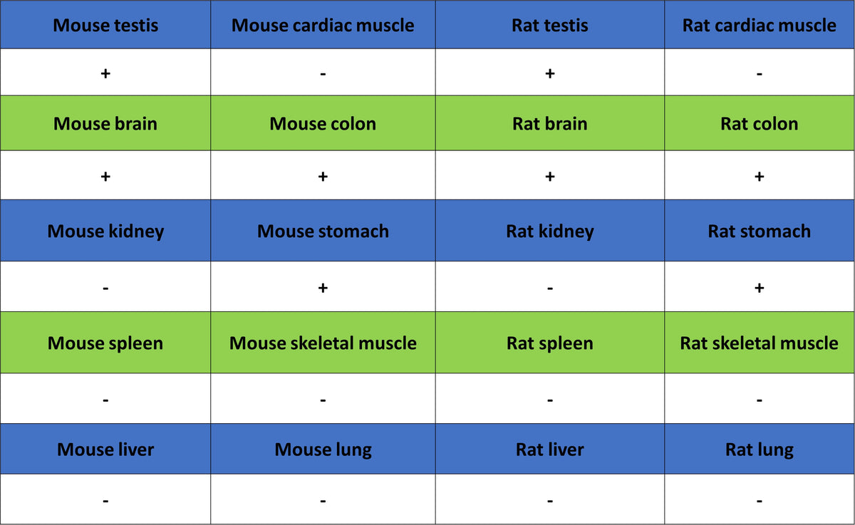
IHC shows positive staining in paraffin-embedded mouse brain. Anti-PGP9.5 antibody was used at 1/2000 dilution, followed by a HRP Polymer for Mouse IgG (ready to use). Counterstained with hematoxylin. Heat mediated antigen retrieval with Tris/EDTA buffer pH9.0 was performed before commencing with IHC staining protocol.
IHC shows positive staining in paraffin-embedded mouse stomach. Anti-PGP9.5 antibody was used at 1/2000 dilution, followed by a HRP Polymer for Mouse IgG (ready to use). Counterstained with hematoxylin. Heat mediated antigen retrieval with Tris/EDTA buffer pH9.0 was performed before commencing with IHC staining protocol.
IHC shows positive staining in paraffin-embedded rat brain. Anti-PGP9.5 antibody was used at 1/2000 dilution, followed by a HRP Polymer for Mouse IgG (ready to use). Counterstained with hematoxylin. Heat mediated antigen retrieval with Tris/EDTA buffer pH9.0 was performed before commencing with IHC staining protocol.
IHC shows positive staining in paraffin-embedded rat pancreas. Anti-PGP9.5 antibody was used at 1/2000 dilution, followed by a HRP Polymer for Mouse IgG (ready to use). Counterstained with hematoxylin. Heat mediated antigen retrieval with Tris/EDTA buffer pH9.0 was performed before commencing with IHC staining protocol.
Immunofluorescence
IF shows positive staining in paraffin-embedded human brain. Anti-PGP9.5 antibody was used at 1/500 dilution (Green) and incubated overnight at 4°C. Goat polyclonal Antibody to mouse IgG - H&L (Alexa Fluor® 488) was used as secondary antibody at 1/1000 dilution. Counterstained with DAPI (Blue). Heat mediated antigen retrieval with EDTA buffer pH9.0 was performed before commencing with IF staining protocol.
IF shows positive staining in paraffin-embedded human kidney. Anti-PGP9.5 antibody was used at 1/500 dilution (Green) and incubated overnight at 4°C. Goat polyclonal Antibody to mouse IgG - H&L (Alexa Fluor® 488) was used as secondary antibody at 1/1000 dilution. Counterstained with DAPI (Blue). Heat mediated antigen retrieval with EDTA buffer pH9.0 was performed before commencing with IF staining protocol.
IF shows positive staining in paraffin-embedded human testis. Anti-PGP9.5 antibody was used at 1/500 dilution (Green) and incubated overnight at 4°C. Goat polyclonal Antibody to mouse IgG - H&L (Alexa Fluor® 488) was used as secondary antibody at 1/1000 dilution. Counterstained with DAPI (Blue). Heat mediated antigen retrieval with EDTA buffer pH9.0 was performed before commencing with IF staining protocol.