WB result of Perilipin-1 Recombinant Rabbit mAb
Primary antibody: Perilipin-1 Recombinant Rabbit mAb at 1/1000 dilution
Lane 1: NIH/3T3 whole cell lysate 20 µg
Lane 2: differentiated 3T3-L whole cell lysate 20 µg
Negative control: NIH/3T3 whole cell lysate
Secondary antibody: Goat Anti-Rabbit IgG (H+L), HRP conjugated at 1/10000 dilution
Predicted MW: 56 kDa
Observed MW: 62 kDa
Perilipin-1 Recombinant Rabbit mAb (S-3093-31)
Perilipin-1 Recombinant Rabbit mAb (S-3093-31)
Price:
Regular price
$100 USD
Regular price
Sale price
$100 USD
Unit price
per
For shipping services or bulk orders, you may request a quotation.
Secure checkout with

View full details
Product Details
Product Details
Product Specification
| Host | Rabbit |
| Antigen | Perilipin-1 |
| Synonyms | Lipid droplet-associated protein; Perilipin A; Peri; Plin; Plin1 |
| Immunogen | Synthetic Peptide |
| Location | Endoplasmic reticulum |
| Accession | Q8CGN5 |
| Clone Number | S-3093-31 |
| Antibody Type | Recombinant mAb |
| Isotype | IgG |
| Application | WB, IHC-P |
| Reactivity | Ms |
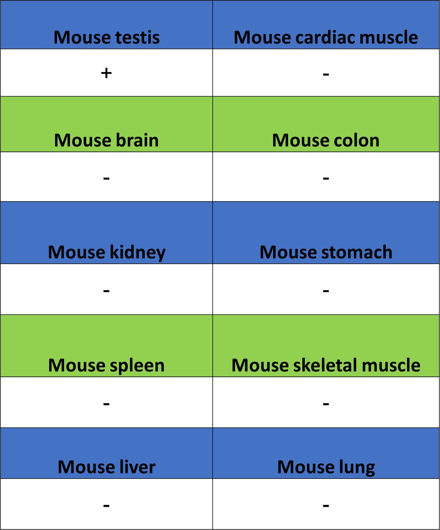
| Positive Sample | differentiated 3T3-L, mouse testis |
| Purification | Protein A |
| Concentration | 0.5 mg/ml |
| Conjugation | Unconjugated |
| Physical Appearance | Liquid |
| Storage Buffer | PBS, 40% Glycerol, 0.05% BSA, 0.02% sodium azide |
| Stability & Storage | 12 months from date of receipt / reconstitution, -20 °C as supplied |
Dilution
| application | dilution | species |
| WB | 1:1000 | Ms |
| IHC-P | 1:2000 | Ms |
Background
Perilipin-1 is a lipid droplet-associated protein encoded by the PLIN1 gene, predominantly expressed in adipocytes of white and brown adipose tissue. This protein consists of 522 amino acids with a molecular weight of approximately 56 kDa, and is localized on the surface of intracellular neutral lipid droplets. It serves as a "molecular coat" on lipid droplets, inhibiting the lipolytic action of hormone-sensitive lipase (HSL) by directly coating the droplets, thereby protecting stored neutral lipids from excessive breakdown. As a key regulator of lipid droplet dynamics and energy homeostasis, phosphorylation of Perilipin-1 is a central mechanism regulating its function—for example, dephosphorylation at Ser511 can modulate lipid droplet size, while BAP31 protein regulates lipid droplet metabolism by influencing its proteasomal degradation. Dysfunction of this protein is closely associated with various human diseases: C-terminal frameshift mutations in the PLIN1 gene can cause familial partial lipodystrophy, characterized by peripheral fat loss, severe hypertriglyceridemia, and insulin resistance in patients. However, studies have also shown that loss-of-function variants in PLIN1 are associated with lower metabolic risk in the population, exhibiting protective features such as elevated HDL cholesterol, reduced triglycerides, and potentially decreased risk of myocardial infarction and hypertension.
Picture
Picture
Western Blot
WB result of Perilipin-1 Recombinant Rabbit mAb
Primary antibody: Perilipin-1 Recombinant Rabbit mAb at 1/1000 dilution
Lane 1: mouse testis lysate 20 µg
Secondary antibody: Goat Anti-Rabbit IgG (H+L), HRP conjugated at 1/10000 dilution
Predicted MW: 56 kDa
Observed MW: 62 kDa
Immunohistochemistry
IHC shows positive staining in paraffin-embedded mouse brown fat. Anti-Perilipin-1 antibody was used at 1/2000 dilution, followed by a HRP Polymer for Rabbit IgG (ready to use). Counterstained with hematoxylin. Heat mediated antigen retrieval with Tris/EDTA buffer pH9.0 was performed before commencing with IHC staining protocol.
IHC shows positive staining in paraffin-embedded mouse white fat. Anti-Perilipin-1 antibody was used at 1/2000 dilution, followed by a HRP Polymer for Rabbit IgG (ready to use). Counterstained with hematoxylin. Heat mediated antigen retrieval with Tris/EDTA buffer pH9.0 was performed before commencing with IHC staining protocol.
IHC shows positive staining in paraffin-embedded mouse testis. Anti-Perilipin-1 antibody was used at 1/2000 dilution, followed by a HRP Polymer for Rabbit IgG (ready to use). Counterstained with hematoxylin. Heat mediated antigen retrieval with Tris/EDTA buffer pH9.0 was performed before commencing with IHC staining protocol.
Negative control: IHC shows negative staining in paraffin-embedded mouse brain. Anti-Perilipin-1 antibody was used at 1/2000 dilution, followed by a HRP Polymer for Rabbit IgG (ready to use). Counterstained with hematoxylin. Heat mediated antigen retrieval with Tris/EDTA buffer pH9.0 was performed before commencing with IHC staining protocol.