WB result of PCK1 Rabbit pAb
Primary antibody: PCK1 Rabbit pAb at 1/1000 dilution
Lane 1: mouse brain lysate 20 µg
Lane 2: mouse adipose lysate 20 µg
Lane 3: mouse kidney lysate 20 µg
Lane 4: mouse liver lysate 20 µg
Secondary antibody: Goat Anti- rabbit IgG (H+L), HRP conjugated at 1/10000 dilution
Predicted MW: 69 kDa
Observed MW: 69 kDa
PCK1 Rabbit Polyclonal Antibody
PCK1 Rabbit Polyclonal Antibody
Price:
Regular price
$100 USD
Regular price
Sale price
$100 USD
Unit price
per
For shipping services or bulk orders, you may request a quotation.
Secure checkout with

View full details
Product Details
Product Details
Product Specification
| Host | Rabbit |
| Antigen | PCK1 |
| Synonyms | Phosphoenolpyruvate carboxykinase, cytosolic [GTP]; PEPCK-C; Serine-protein kinase PCK1; PEPCK1 |
| Immunogen | Synthetic Peptide |
| Location | Cytoplasm, Endoplasmic reticulum |
| Accession | P35558 |
| Antibody Type | Polyclonal antibody |
| Isotype | IgG |
| Application | WB, IHC-P, IF |
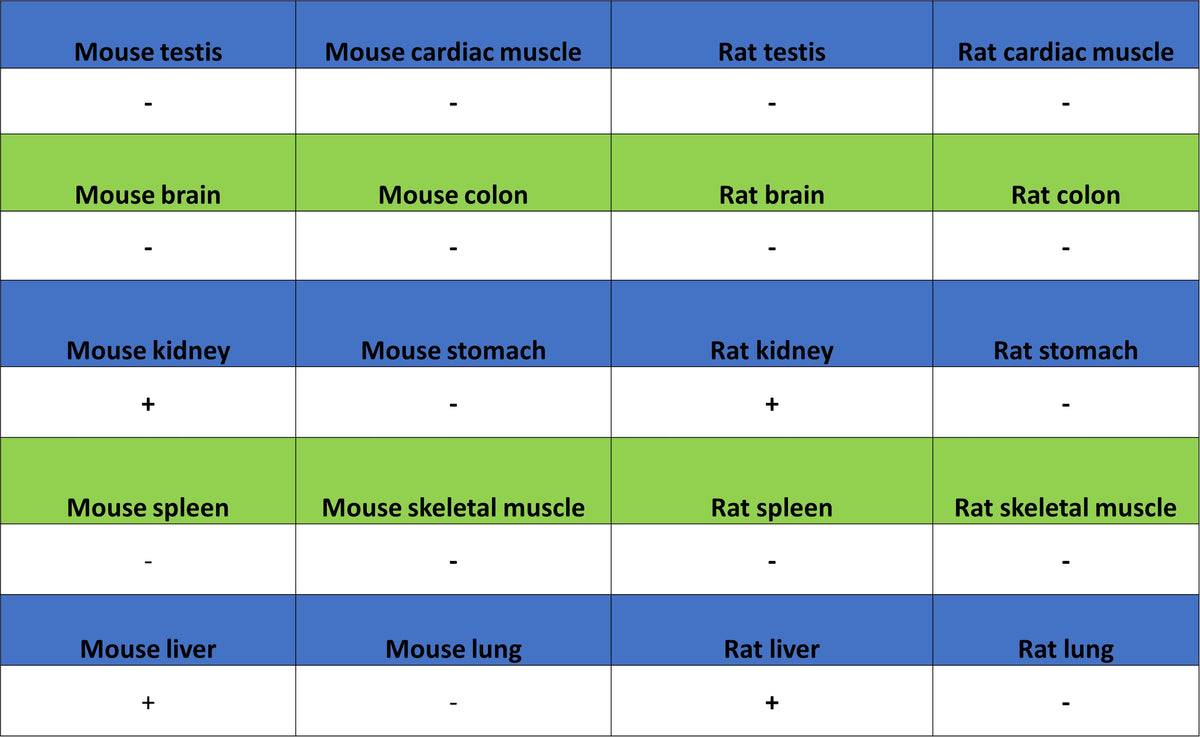
| Reactivity | Ms, Rt |
| Purification | Immunogen Affinity |
| Concentration | 0.5 mg/ml |
| Conjugation | Unconjugated |
| Physical Appearance | Liquid |
| Storage Buffer | PBS, 40% Glycerol, 0.05% BSA, 0.02% sodium azide |
| Stability & Storage | 12 months from date of receipt / reconstitution, -20 °C as supplied |
Dilution
| application | dilution | species |
| WB | 1:1000-1:5000 | Ms, Rt |
| IHC-P | 1:500 | Ms, Rt |
| IF | 1:500 | Ms, Rt |
Background
PCK1, also known as phosphoenolpyruvate carboxykinase 1, is a crucial enzyme involved in gluconeogenesis and the conversion of oxaloacetate to phosphoenolpyruvate. It plays a significant role in maintaining glucose homeostasis by facilitating the production of glucose from non-carbohydrate sources. This enzyme is primarily expressed in tissues such as the liver and kidneys, where gluconeogenesis predominantly occurs. Abnormal regulation or activity of PCK1 has been implicated in metabolic disorders, including diabetes.
Picture
Picture
Western Blot
WB result of PCK1 Rabbit pAb
Primary antibody: PCK1 Rabbit pAb at 1/1000 dilution
Lane 1: rat brain lysate 20 µg
Lane 2: rat kidney lysate 20 µg
Lane 3: rat liver lysate 20 µg
Secondary antibody: Goat Anti- rabbit IgG (H+L), HRP conjugated at 1/10000 dilution
Predicted MW: 69 kDa
Observed MW: 69 kDa
Immunohistochemistry
IHC shows positive staining in paraffin-embedded mouse kidney. Anti-NTSR1 antibody was used at 1/4000 dilution, followed by a HRP Polymer for Rabbit IgG (ready to use). Counterstained with hematoxylin. Heat mediated antigen retrieval with Tris/EDTA buffer pH9.0 was performed before commencing with IHC staining protocol.
IHC shows positive staining in paraffin-embedded mouse liver. Anti-NTSR1 antibody was used at 1/4000 dilution, followed by a HRP Polymer for Rabbit IgG (ready to use). Counterstained with hematoxylin. Heat mediated antigen retrieval with Tris/EDTA buffer pH9.0 was performed before commencing with IHC staining protocol.
IHC shows positive staining in paraffin-embedded rat kidney. Anti-NTSR1 antibody was used at 1/4000 dilution, followed by a HRP Polymer for Rabbit IgG (ready to use). Counterstained with hematoxylin. Heat mediated antigen retrieval with Tris/EDTA buffer pH9.0 was performed before commencing with IHC staining protocol.
Immunofluorescence
IF shows positive staining in paraffin-embedded mouse kidney. Anti-PCK1 antibody was used at 1/500 dilution (Green) and incubated overnight at 4°C. Goat polyclonal Antibody to Rabbit IgG - H&L (Alexa Fluor® 488) was used as secondary antibody at 1/1000 dilution. Counterstained with DAPI (Blue). Heat mediated antigen retrieval with EDTA buffer pH9.0 was performed before commencing with IF staining protocol.
IF shows positive staining in paraffin-embedded rat liver. Anti-PCK1 antibody was used at 1/500 dilution (Green) and incubated overnight at 4°C. Goat polyclonal Antibody to Rabbit IgG - H&L (Alexa Fluor® 488) was used as secondary antibody at 1/1000 dilution. Counterstained with DAPI (Blue). Heat mediated antigen retrieval with EDTA buffer pH9.0 was performed before commencing with IF staining protocol.