WB result of ITPR1 Recombinant Rabbit mAb
Primary antibody: ITPR1 Recombinant Rabbit mAb at 1/1000 dilution
Lane 1: mouse cerebellum lysate 20 μg
Secondary antibody: Goat Anti-Rabbit IgG (H+L), HRP conjugated at 1/10000 dilution
Predicted MW: 314 kDa
Observed MW: 290 kDa
This blot was developed with high sensitivity substrate
ITPR1 Recombinant Rabbit mAb (S-4530-5)
ITPR1 Recombinant Rabbit mAb (S-4530-5)
Price:
Regular price
$100 USD
Regular price
Sale price
$100 USD
Unit price
per
For shipping services or bulk orders, you may request a quotation.
Secure checkout with

View full details
Product Details
Product Details
Product Specification
| Host | Rabbit |
| Antigen | ITPR1 |
| Synonyms | Inositol 1,4,5-trisphosphate-gated calcium channel ITPR1; IP3 receptor isoform 1 (IP3R 1; InsP3R1); Inositol 1,4,5 trisphosphate receptor; Inositol 1,4,5-trisphosphate receptor type 1; Type 1 inositol 1,4,5-trisphosphate receptor (Type 1 InsP3 receptor); INSP3R1 |
| Immunogen | Synthetic Peptide |
| Location | Cytoplasm, Endoplasmic reticulum |
| Accession | Q14643 |
| Clone Number | S-4530-5 |
| Antibody Type | Recombinant mAb |
| Isotype | IgG |
| Application | WB, IHC-P, IF |
| Reactivity | Hu, Ms, Rt |
| Positive Sample | mouse cerebellum, rat cerebellum |
| Predicted Reactivity | Bv |
| Purification | Protein A |
| Concentration | 0.5 mg/ml |
| Conjugation | Unconjugated |
| Physical Appearance | Liquid |
| Storage Buffer | PBS, 40% Glycerol, 0.05% BSA, 0.02% sodium azide |
| Stability & Storage | 12 months from date of receipt / reconstitution, -20 °C as supplied |
Dilution
| application | dilution | species |
| WB | 1:1000 | Ms, Rt |
| IHC-P | 1:2000 | Hu, Ms, Rt |
| IF | 1:500 | Hu, Ms, Rt |
Background
ITPR1 (inositol 1,4,5-trisphosphate receptor type 1) is a ligand-gated calcium release channel located on the endoplasmic reticulum membrane, encoded by the ITPR1 gene, which serves as a core hub in intracellular calcium signaling by converting extracellular signals into calcium release from the endoplasmic reticulum calcium store into the cytoplasm through binding the second messenger inositol 1,4,5-trisphosphate (IP3). This protein has a large molecular structure comprising an N-terminal regulatory domain that binds IP3, a C-terminal transmembrane domain at the core of the channel, and multiple signaling regulatory sites distributed throughout the molecule, enabling its function to be precisely regulated by IP3 concentration, cytoplasmic calcium, ATP, redox status, and various phosphorylation modifications. In the nervous system, ITPR1 is highly enriched in neurons such as Purkinje cells, and the calcium signals it mediates are essential for regulating synaptic plasticity, dendritic development, and neuronal excitability; consequently, loss-of-function mutations or autoantibody-mediated dysfunction of this gene have been identified as the cause of various neurological disorders, including spinocerebellar ataxia type 15 (SCA15), SCA29, Gillespie syndrome, and autoimmune cerebellar ataxia. Beyond its neurological functions, ITPR1 occupies a central position in the global regulation of intracellular calcium homeostasis, and its functional abnormalities are closely associated with cardiac diseases, severe acute pancreatitis, immune cell activation, and invasion and metastasis in various cancers (such as glioblastoma and breast cancer); meanwhile, the interaction of this channel with anti-apoptotic proteins (such as the Bcl-2 family) also makes it a critical node in regulating cell fate decisions, highlighting its great potential as a therapeutic target for multiple diseases.
Picture
Picture
Western Blot
WB result of ITPR1 Recombinant Rabbit mAb
Primary antibody: ITPR1 Recombinant Rabbit mAb at 1/1000 dilution
Lane 1: rat cerebellum lysate 20 μg
Secondary antibody: Goat Anti-Rabbit IgG (H+L), HRP conjugated at 1/10000 dilution
Predicted MW: 314 kDa
Observed MW: 290 kDa
This blot was developed with high sensitivity substrate
Immunohistochemistry
IHC shows positive staining in paraffin-embedded human cerebellum. Anti-ITPR1 antibody was used at 1/2000 dilution, followed by a HRP Polymer for Rabbit IgG (ready to use). Counterstained with hematoxylin. Heat mediated antigen retrieval with Tris/EDTA buffer pH9.0 was performed before commencing with IHC staining protocol.
Negative control: IHC shows negative staining in paraffin-embedded human colon. Anti-ITPR1 antibody was used at 1/2000 dilution, followed by a HRP Polymer for Rabbit IgG (ready to use). Counterstained with hematoxylin. Heat mediated antigen retrieval with Tris/EDTA buffer pH9.0 was performed before commencing with IHC staining protocol.
Negative control: IHC shows negative staining in paraffin-embedded human hepatocellular carcinoma. Anti-ITPR1 antibody was used at 1/2000 dilution, followed by a HRP Polymer for Rabbit IgG (ready to use). Counterstained with hematoxylin. Heat mediated antigen retrieval with Tris/EDTA buffer pH9.0 was performed before commencing with IHC staining protocol.
IHC shows positive staining in paraffin-embedded rat cerebellum. Anti-ITPR1 antibody was used at 1/2000 dilution, followed by a HRP Polymer for Rabbit IgG (ready to use). Counterstained with hematoxylin. Heat mediated antigen retrieval with Tris/EDTA buffer pH9.0 was performed before commencing with IHC staining protocol.
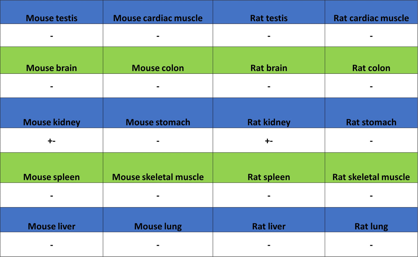
Negative control: IHC shows negative staining in paraffin-embedded mouse testis. Anti-ITPR1 antibody was used at 1/2000 dilution, followed by a HRP Polymer for Rabbit IgG (ready to use). Counterstained with hematoxylin. Heat mediated antigen retrieval with Tris/EDTA buffer pH9.0 was performed before commencing with IHC staining protocol.
IHC shows positive staining in paraffin-embedded rat cerebellum. Anti-ITPR1 antibody was used at 1/2000 dilution, followed by a HRP Polymer for Rabbit IgG (ready to use). Counterstained with hematoxylin. Heat mediated antigen retrieval with Tris/EDTA buffer pH9.0 was performed before commencing with IHC staining protocol.
Negative control: IHC shows negative staining in paraffin-embedded rat spleen. Anti-ITPR1 antibody was used at 1/2000 dilution, followed by a HRP Polymer for Rabbit IgG (ready to use). Counterstained with hematoxylin. Heat mediated antigen retrieval with Tris/EDTA buffer pH9.0 was performed before commencing with IHC staining protocol.
Immunofluorescence
IF shows positive staining in paraffin-embedded human cerebellum. Anti-ITPR1 antibody was used at 1/500 dilution (Green) and incubated overnight at 4°C. Goat polyclonal Antibody to Rabbit IgG - H&L (Alexa Fluor® 488) was used as secondary antibody at 1/1000 dilution. Counterstained with DAPI (Blue). Heat mediated antigen retrieval with EDTA buffer pH9.0 was performed before commencing with IF staining protocol.
IF shows positive staining in paraffin-embedded mouse cerebellum. Anti-ITPR1 antibody was used at 1/500 dilution (Green) and incubated overnight at 4°C. Goat polyclonal Antibody to Rabbit IgG - H&L (Alexa Fluor® 488) was used as secondary antibody at 1/1000 dilution. Counterstained with DAPI (Blue). Heat mediated antigen retrieval with EDTA buffer pH9.0 was performed before commencing with IF staining protocol.
IF shows positive staining in paraffin-embedded rat cerebellum. Anti-ITPR1 antibody was used at 1/500 dilution (Green) and incubated overnight at 4°C. Goat polyclonal Antibody to Rabbit IgG - H&L (Alexa Fluor® 488) was used as secondary antibody at 1/1000 dilution. Counterstained with DAPI (Blue). Heat mediated antigen retrieval with EDTA buffer pH9.0 was performed before commencing with IF staining protocol.