WB result of IFIT2 Recombinant Rabbit mAb
Primary antibody: IFIT2 Recombinant Rabbit mAb at 1/1000 dilution
Lane 1: HCT 116 whole cell lysate 20 μg
Lane 2: Raji whole cell lysate 20 μg
Lane 3: A431 whole cell lysate 20 μg
Low expression control: HCT 116 whole cell lysate, Raji whole cell lysate
Secondary antibody: Goat Anti-Rabbit IgG (H+L), HRP conjugated at 1/10000 dilution
Predicted MW: 55 kDa
Observed MW: 55 kDa
This blot was developed with high sensitivity substrate
IFIT2 Recombinant Rabbit mAb (S-6029)
IFIT2 Recombinant Rabbit mAb (S-6029)
Price:
Regular price
$100 USD
Regular price
Sale price
$100 USD
Unit price
per
For shipping services or bulk orders, you may request a quotation.
Secure checkout with

View full details
Product Details
Product Details
Product Specification
| Host | Rabbit |
| Antigen | IFIT2 |
| Synonyms | Interferon-induced protein with tetratricopeptide repeats 2; ISG-54 K; Interferon-induced 54 kDa protein (IFI-54K; P54); CIG-42; G10P2; IFI54; ISG54 |
| Location | Cytoplasm, Endoplasmic reticulum |
| Accession | P09913 |
| Clone Number | S-6029 |
| Antibody Type | Recombinant mAb |
| Isotype | IgG |
| Application | WB, IHC-P |
| Reactivity | Hu |
| Positive Sample | A431 |
| Purification | Protein A |
| Concentration | 0.5 mg/ml |
| Conjugation | Unconjugated |
| Physical Appearance | Liquid |
| Storage Buffer | PBS, 40% Glycerol, 0.05% BSA, 0.02% sodium azide |
| Stability & Storage | 12 months from date of receipt / reconstitution, -20 °C as supplied |
Dilution
| application | dilution | species |
| WB | 1:1000 | Hu |
| IHC-P | 1:500 | Hu |
Background
IFIT2 (interferon-induced protein with tetratricopeptide repeats 2, also known as ISG54 or GARG-39) is an interferon-stimulated gene (ISG) encoded by the IFIT2 gene, belonging to the IFIT protein family, whose core function is to serve as a key effector in intracellular viral recognition and restriction. This protein contains multiple tetratricopeptide repeat (TPR) domains, which confer its unique ability to interact with proteins, enabling it to bind and neutralize key components of viral invasion from various viruses, such as recognizing viral mRNA lacking 2'-O-methylated cap structures or binding to viral RNA polymerase complexes, thereby directly inhibiting viral mRNA translation initiation or replication processes. Beyond its classical antiviral functions, accumulating evidence has revealed the complex roles of IFIT2 in tumor biology: on one hand, it exhibits tumor-suppressive properties in cancers such as gastric cancer and melanoma by promoting the resolution of cancer-associated inflammation or enhancing immune surveillance, with its high expression typically correlating with better patient prognosis; on the other hand, in certain tumor types or specific microenvironments, IFIT2 may also be hijacked by tumor cells to promote metastasis and chemoresistance through regulating epithelial-mesenchymal transition (EMT) or activating specific survival pathways, and this functional duality makes it a hotspot molecule in cancer research. In addition, IFIT2 expression is finely regulated by both interferon and non-interferon signals, and its gene polymorphisms are associated with various autoimmune diseases and viral susceptibility, while significant expression changes have also been observed in non-infectious stress responses such as ischemia-reperfusion injury, indicating broader biological functions of this protein beyond immune defense.
Picture
Picture
Western Blot
Immunohistochemistry
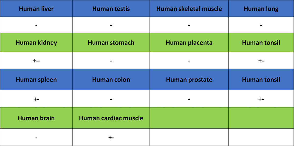
IHC shows positive staining in paraffin-embedded human kidney. Anti-IFIT2 antibody was used at 1/500 dilution, followed by a HRP Polymer for Rabbit IgG (ready to use). Counterstained with hematoxylin. Heat mediated antigen retrieval with Tris/EDTA buffer pH9.0 was performed before commencing with IHC staining protocol.
IHC shows positive staining in paraffin-embedded human cervical cancer. Anti-IFIT2 antibody was used at 1/500 dilution, followed by a HRP Polymer for Rabbit IgG (ready to use). Counterstained with hematoxylin. Heat mediated antigen retrieval with Tris/EDTA buffer pH9.0 was performed before commencing with IHC staining protocol.
Negative control: IHC shows negative staining in paraffin-embedded human skeletal muscle. Anti-IFIT2 antibody was used at 1/500 dilution, followed by a HRP Polymer for Rabbit IgG (ready to use). Counterstained with hematoxylin. Heat mediated antigen retrieval with Tris/EDTA buffer pH9.0 was performed before commencing with IHC staining protocol.